第33章動物の体:基本的な形態と機能
Fibrous Connective Tissue線維性結合組織
Fibrous connective tissues contain large amounts of collagen fibers and few cells or matrix material. The fibers can be arranged irregularly or regularly with the strands lined up in parallel. Irregularly arranged fibrous connective tissues are found in areas of the body where stress occurs from all directions, such as the dermis of the skin. Regular fibrous connective tissue, shown in Figure 33.13, is found in tendons (which connect muscles to bones) and ligaments (which connect bones to bones).
線維性結合組織には、多量の膠原線維と、少量の細胞や基質が含まれる。線維は不規則に並ぶことも、平行な束として規則的に並ぶこともある。不規則な配列の線維性結合組織は、皮膚の真皮のように、あらゆる方向から力がかかる場所に見られる。図33.13の規則的な線維性結合組織は、筋肉と骨をつなぐ腱や、骨と骨をつなぐ靱帯に見られる。
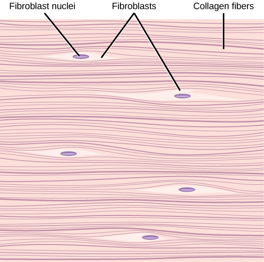
Figure 33.13図 33.13
Fibrous connective tissue from the tendon has strands of collagen fibers lined up in parallel.
腱の線維性結合組織では、膠原線維の束が平行に並ぶ。
Cartilage軟骨
Cartilage is a connective tissue with a large amount of the matrix of variable composition. The cells, called chondrocytes, make the matrix and fibers of the tissue. Chondrocytes are found in spaces within the tissue called lacunae.
軟骨は、組成の異なる多量の細胞外基質をもつ結合組織である。軟骨細胞と呼ばれる細胞が、組織の基質と線維を作る。軟骨細胞は、組織内の軟骨小腔という空間にある。
A cartilage with few collagen and elastic fibers is hyaline cartilage, illustrated in Figure 33.14. The lacunae are randomly scattered throughout the tissue and the matrix takes on a milky or scrubbed appearance with routine histological stains. Sharks have cartilaginous skeletons, as does nearly the entire human skeleton during a specific pre-birth developmental stage. A remnant of this cartilage persists in the outer portion of the human nose. Hyaline cartilage is also found at the ends of long bones, reducing friction and cushioning the articulations of these bones.
英語のヒント
膠原線維と弾性線維が少ない軟骨が、図33.14に示す硝子軟骨である。小腔は組織全体に不規則に散らばり、通常の組織染色では、基質が乳白色やすりガラスのように見える。サメは軟骨性の骨格をもつ。また、ヒトの骨格も出生前の特定の発生段階では、ほぼ全体が軟骨でできている。その軟骨の一部は、ヒトの鼻の外側部分に残る。硝子軟骨は長骨の端にもあり、摩擦を減らし、骨が接する関節部分の衝撃を和らげる。
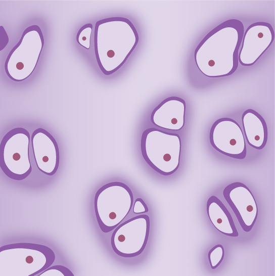
Figure 33.14図 33.14
Hyaline cartilage consists of a matrix with cells called chondrocytes embedded in it. The chondrocytes exist in cavities in the matrix called lacunae.
硝子軟骨は、軟骨細胞が埋め込まれた細胞外基質からなる。軟骨細胞は、基質内の小腔と呼ばれる空洞にある。
Elastic cartilage has a large amount of elastic fibers, giving it tremendous flexibility. The ears of most vertebrate animals contain this cartilage as do portions of the larynx, or voice box. Fibrocartilage contains a large amount of collagen fibers, giving the tissue tremendous strength. Fibrocartilage comprises the intervertebral discs in vertebrate animals. Hyaline cartilage found in movable joints such as the knee and shoulder becomes damaged as a result of age or trauma. Damaged hyaline cartilage is replaced by fibrocartilage and results in the joints becoming “stiff.”
弾性軟骨は多量の弾性線維をもち、非常に柔軟である。大部分の脊椎動物の耳と、発声器官である喉頭の一部に含まれる。線維軟骨は多量の膠原線維を含み、きわめて強い組織となる。脊椎動物の椎間板を構成するのが線維軟骨である。膝や肩のような可動性の関節にある硝子軟骨は、加齢や外傷によって損傷する。損傷した硝子軟骨は線維軟骨に置き換わり、関節が「硬く」なる。
Bone骨
Bone, or osseous tissue, is a connective tissue that has a large amount of two different types of matrix material. The organic matrix is similar to the matrix material found in other connective tissues, including some amount of collagen and elastic fibers. This gives strength and flexibility to the tissue. The inorganic matrix consists of mineral salts—mostly calcium salts—that give the tissue hardness. Without adequate organic material in the matrix, the tissue breaks; without adequate inorganic material in the matrix, the tissue bends.
骨、すなわち骨組織は、異なる2種類の基質成分を大量に含む結合組織である。有機性の基質は、ほかの結合組織の基質に似ており、ある程度の膠原線維や弾性線維を含む。これが組織に強さと柔軟性を与える。無機性の基質は、主にカルシウム塩からなる無機塩類でできており、組織に硬さを与える。基質に十分な有機物がなければ組織は折れ、十分な無機物がなければ曲がる。
There are three types of cells in bone: osteoblasts, osteocytes, and osteoclasts. Osteoblasts are active in making bone for growth and remodeling. Osteoblasts deposit bone material into the matrix and, after the matrix surrounds them, they continue to live, but in a reduced metabolic state as osteocytes. Osteocytes are found in lacunae of the bone. Osteoclasts are active in breaking down bone for bone remodeling, and they provide access to calcium stored in tissues. Osteoclasts are usually found on the surface of the tissue.
英語のヒント
骨には、骨芽細胞、骨細胞、破骨細胞という3種類の細胞がある。骨芽細胞は、成長やリモデリングのための骨形成を活発に行う。基質へ骨の成分を沈着させ、やがて基質に囲まれると、代謝活動を抑えた骨細胞として生き続ける。骨細胞は骨の小腔内にある。破骨細胞は、骨のリモデリングのために骨を活発に分解し、組織に蓄えられたカルシウムを利用できるようにする。通常、組織の表面に見られる。
Bone can be divided into two types: compact and spongy. Compact bone is found in the shaft (or diaphysis) of a long bone and the surface of the flat bones, while spongy bone is found in the end (or epiphysis) of a long bone. Compact bone is organized into subunits called osteons, as illustrated in Figure 33.15. A blood vessel and a nerve are found in the center of the structure within the Haversian canal, with radiating circles of lacunae around it known as lamellae. The wavy lines seen between the lacunae are microchannels called canaliculi; they connect the lacunae to aid diffusion between the cells. Spongy bone is made of tiny plates called trabeculae; these plates serve as struts to give the spongy bone strength. Over time, these plates can break causing the bone to become less resilient. Bone tissue forms the internal skeleton of vertebrate animals, providing structure to the animal and points of attachment for tendons.
英語のヒント
骨は緻密骨と海綿骨に分けられる。緻密骨は長骨の骨幹と扁平骨の表面にあり、海綿骨は長骨の端、すなわち骨端にある。図33.15のように、緻密骨はオステオンという単位で組織化されている。中央のハバース管には血管と神経があり、その周囲に、層板と呼ばれる同心円状の小腔の配列が広がる。小腔の間に見える波状の線は、骨細管という細い通路である。小腔同士をつないで、細胞間の拡散を助ける。海綿骨は骨梁という小さな板からなり、それらが支柱として働いて強さを与える。時間がたつと板が折れ、骨のしなやかな強さが低下することがある。骨組織は脊椎動物の内骨格を作り、体を支える構造と、腱の付着点を与える。
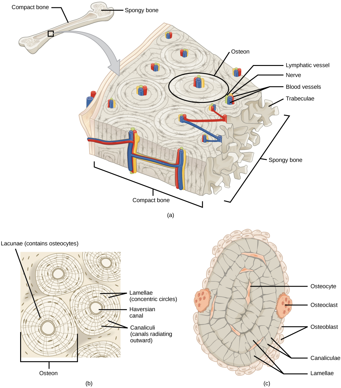


Figure 33.15図 33.15
(a) Compact bone is a dense matrix on the outer surface of bone. Spongy bone, inside the compact bone, is porous with web-like trabeculae. (b) Compact bone is organized into rings called osteons. Blood vessels, nerves, and lymphatic vessels are found in the central Haversian canal. Rings of lamellae surround the Haversian canal. Between the lamellae are cavities called lacunae. Canaliculi are microchannels connecting the lacunae together. (c) Osteoblasts surround the exterior of the bone. Osteoclasts bore tunnels into the bone and osteocytes are found in the lacunae.
英語のヒント
(a)緻密骨は骨の外表面にある密な基質である。その内側の海綿骨は、網目状の骨梁をもつ多孔性の構造である。(b)緻密骨は、オステオンという輪状の単位に組織化される。中央のハバース管には、血管、神経、リンパ管がある。層板の輪がハバース管を囲み、その間に小腔がある。骨細管は、小腔同士をつなぐ微細な通路である。(c)骨芽細胞は骨の外側を囲む。破骨細胞は骨にトンネルを掘り、骨細胞は小腔内にある。