第21章ウイルス
21.1 Viral Evolution, Morphology, and Classification21.1 ウイルスの進化・形態・分類
Learning Objectives学習目標
By the end of this section, you will be able to do the following:
この節を終えると、次のことができるようになる。
- Describe how viruses were first discovered and how they are detectedウイルスが最初に発見された経緯と、その検出方法を説明する。
- Discuss three hypotheses about how viruses evolvedウイルスの進化に関する3つの仮説を論じる。
- Describe the general structure of a virusウイルスの一般的な構造を説明する。
- Recognize the basic shapes of virusesウイルスの基本的な形を識別する。
- Understand past and emerging classification systems for virusesウイルスの過去の分類体系と、新たに生まれつつある分類体系を理解する。
- Describe the basis for the Baltimore classification systemボルティモア分類の基準を説明する。
Viruses are diverse entities: They vary in structure, methods of replication, and the hosts they infect. Nearly all forms of life—from prokaryotic bacteria and archaeans, to eukaryotes such as plants, animals, and fungi—have viruses that infect them. While most biological diversity can be understood through evolutionary history (such as how species have adapted to changing environmental conditions and how different species are related to one another through common descent), much about virus origins and evolution remains unknown.
英語のヒント
ウイルスは多様な存在であり、構造、複製の方法、感染する宿主が異なる。原核生物の細菌や古細菌から、植物、動物、菌類などの真核生物まで、ほぼすべての生命の型に、それらへ感染するウイルスがある。生物の多様性の大部分は、種が変化する環境条件にどう適応してきたか、異なる種が共通の由来によってどうつながるか、といった進化史を通して理解できる。しかし、ウイルスの起源と進化については、今なお多くが分かっていない。
Discovery and Detection発見と検出
Viruses were first discovered after the development of a porcelain filter—the Chamberland-Pasteur filter—that could remove all bacteria visible in the microscope from any liquid sample. In 1886, Adolph Meyer demonstrated that a disease of tobacco plants—tobacco mosaic disease—could be transferred from a diseased plant to a healthy one via liquid plant extracts. In 1892, Dmitri Ivanowski showed that this disease could be transmitted in this way even after the Chamberland-Pasteur filter had removed all viable bacteria from the extract. Still, it was many years before it was proved that these “filterable” infectious agents were not simply very small bacteria but were a new type of very small, disease-causing particle.
英語のヒント
ウイルスが初めて発見されたのは、液体試料から、顕微鏡で見えるすべての細菌を取り除ける磁器製フィルター、シャンベラン=パスツール濾過器が開発された後だった。1886年、アドルフ・マイヤーは、タバコの病気であるタバコモザイク病が、植物の液状抽出物を介して、病気の植物から健康な植物へ移ることを示した。1892年、ドミトリ・イワノフスキーは、この濾過器によって抽出液から生きた細菌をすべて除いた後でも、同じ方法で病気が伝わることを示した。それでも、これらの「濾過性」の感染因子が単に非常に小さな細菌ではなく、新しい種類のごく小さな病原性粒子であると証明されるまでには、さらに長い年月がかかった。
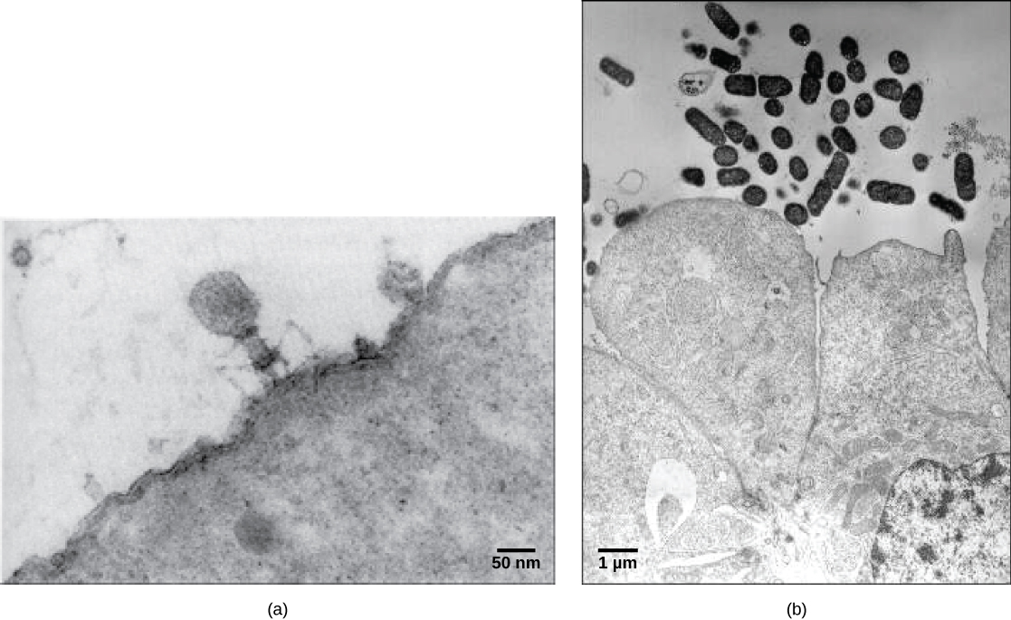
Most virions, or single virus particles, are very small, about 20 to 250 nanometers in diameter. However, some recently discovered viruses from amoebae range up to 1000 nm in diameter. With the exception of large virions, like the poxvirus and other large DNA viruses, viruses cannot be seen with a light microscope. It was not until the development of the electron microscope in the late 1930s that scientists got their first good view of the structure of the tobacco mosaic virus (TMV) (Figure 21.1), discussed above, and other viruses (Figure 21.2). The surface structure of virions can be observed by both scanning and transmission electron microscopy, whereas the internal structures of the virus can only be observed in images from a transmission electron microscope. The use of electron microscopy and other technologies has allowed for the discovery of many viruses of all types of living organisms.
英語のヒント
大半のビリオン、すなわち個々のウイルス粒子は、直径約20~250ナノメートルと非常に小さい。ただし、近年アメーバから発見されたウイルスには、直径1000 nmに達するものもある。ポックスウイルスや他の大型DNAウイルスのような大きなビリオンを除き、光学顕微鏡ではウイルスを見ることはできない。1930年代後半に電子顕微鏡が開発されて初めて、科学者は先に述べたタバコモザイクウイルス(TMV、図21.1)や他のウイルス(図21.2)の構造をはっきり観察できた。ビリオンの表面構造は走査型と透過型のどちらの電子顕微鏡でも観察できるが、内部構造は透過型電子顕微鏡の画像でしか見られない。電子顕微鏡などの技術を用いることで、あらゆる種類の生物に感染する多数のウイルスが発見された。
OpenStax, Rice University; Biology 2e. (credit a: modification of work by U.S. Dept. of Energy, Office of Science, LBL, PBD; credit b: modification of work by J.P. Nataro and S. Sears, unpub. data, CDC; scale-bar data from Matt Russell) · CC BY 4.0 · Source出典

Figure 21.2図 21.2
Most virus particles are visible only by electron microscopy. In these transmission electron micrographs, (a) a virus is as dwarfed by the bacterial cell it infects, as (b) these E. coli cells are dwarfed by cultured colon cells. (credit a: modification of work by U.S. Dept. of Energy, Office of Science, LBL, PBD; credit b: modification of work by J.P. Nataro and S. Sears, unpub. data, CDC; scale-bar data from Matt Russell)
英語のヒント
大半のウイルス粒子は、電子顕微鏡でしか見えない。これらの透過型電子顕微鏡写真では、(a)ウイルスは感染先の細菌細胞と比べて非常に小さく、同じように(b)大腸菌細胞は培養した結腸細胞と比べて非常に小さい。(a:米国エネルギー省科学局、LBL、PBDの作品を改変。b:J.P. NataroとS. Sears、未発表データ、CDCの作品を改変。スケールバーのデータ:Matt Russell)
Evolution of Virusesウイルスの進化
Although biologists have a significant amount of knowledge about how present-day viruses mutate and adapt, much less is known about how viruses originated in the first place. When exploring the evolutionary history of most organisms, scientists can look at fossil records and similar historic evidence. However, viruses do not fossilize, as far as we know, so researchers must extrapolate from investigations of how today’s viruses evolve and by using biochemical and genetic information to create speculative virus histories.
英語のヒント
生物学者は、現代のウイルスがどう変異し適応するかについて多くを知っている。しかし、ウイルスがそもそもどのように生じたかについて分かっていることは、はるかに少ない。大半の生物の進化史を調べる場合、科学者は化石記録や類似の歴史的証拠を参照できる。しかし、知られている限りウイルスは化石にならない。そのため研究者は、現在のウイルスの進化の調査から過去を推し量り、生化学的・遺伝的情報も使って、ウイルスの歴史を推測しなければならない。
Most scholars agree that viruses don’t have a single common ancestor, nor is there a single reasonable hypothesis about virus origins. There are current evolutionary scenarios that may explain the origin of viruses. One such hypothesis, the “devolution” or the regressive hypothesis, suggests that viruses evolved from free-living cells, or from intracellular prokaryotic parasites. However, many components of how this process might have occurred remain a mystery. A second hypothesis, the escapist or the progressive hypothesis, suggests that viruses originated from RNA and DNA molecules, or self-replicating entities similar to transposons or other mobile genetic elements, that escaped from a host cell with the ability to enter another. A third hypothesis, the virus first hypothesis, suggests that viruses may have been the first self-replicating entities before the first cells. In all cases, viruses are probably continuing to evolve along with the cells on which they rely on as hosts.
英語のヒント
大半の研究者は、ウイルスに単一の共通祖先はなく、その起源について唯一の妥当な仮説もないという点で一致している。現在、ウイルスの起源を説明しうる進化の筋書きがいくつかある。その1つである「退化」仮説、または退行仮説では、ウイルスは自由生活性の細胞、あるいは細胞内に寄生する原核生物から進化したと考える。しかし、その過程がどう起こったかには、なお多くの謎がある。第2の逃避仮説、または進行仮説では、ウイルスはRNAやDNA分子、あるいはトランスポゾンなどの可動性遺伝因子に似た自己複製体に由来すると考える。それらが、別の細胞に入る能力を伴って宿主細胞から抜け出したというのである。第3のウイルス先行仮説では、ウイルスは最初の細胞より前に存在した、最初の自己複製体だった可能性があるとする。いずれの場合も、ウイルスは宿主として依存する細胞とともに、今も進化し続けているのだろう。
As technology advances, scientists may develop and refine additional hypotheses to explain the origins of viruses. The emerging field called virus molecular systematics attempts to do just that through comparisons of sequenced genetic material. These researchers hope one day to better understand the origin of viruses—a discovery that could lead to advances in the treatments for the ailments they produce.
英語のヒント
技術の進歩に伴い、科学者はウイルスの起源を説明する追加の仮説を作り、洗練させていくだろう。新たに生まれつつあるウイルス分子系統学という分野は、配列が決定された遺伝物質を比較することで、まさにそれを目指している。研究者は、いつかウイルスの起源をよりよく理解できることを願っている。その発見は、ウイルスが起こす病気の治療の進歩につながるかもしれない。