Unlike nearly all living organisms that use DNA as their genetic material, viruses may use either DNA or RNA. The virus core contains the genome—the total genetic content of the virus. Viral genomes tend to be small, containing only those genes that encode proteins which the virus cannot get from the host cell. This genetic material may be single- or double-stranded. It may also be linear or circular. While most viruses contain a single nucleic acid, others have genomes divided into several segments. The RNA genome of the influenza virus is segmented, which contributes to its variability and continuous evolution, and explains why it is difficult to develop a vaccine against it.
In DNA viruses, the viral DNA directs the host cell’s replication proteins to synthesize new copies of the viral genome and to transcribe and translate that genome into viral proteins. Human diseases caused by DNA viruses include chickenpox, hepatitis B, and adenoviruses. Sexually transmitted DNA viruses include the herpes virus and the human papilloma virus (HPV), which has been associated with cervical cancer and genital warts.
RNA viruses contain only RNA as their genetic material. To replicate their genomes in the host cell, the RNA viruses must encode their own enzymes that can replicate RNA into RNA or, in the retroviruses, into DNA. These RNA polymerase enzymes are more likely to make copying errors than DNA polymerases, and therefore often make mistakes during transcription. For this reason, mutations in RNA viruses occur more frequently than in DNA viruses. This causes them to change and adapt more rapidly to their host. Human diseases caused by RNA viruses include influenza, hepatitis C, measles, and rabies. The HIV virus, which is sexually transmitted, is an RNA retrovirus.
Because most viruses probably evolved from different ancestors, the systematic methods that scientists have used to classify prokaryotic and eukaryotic cells are not very useful. If viruses represent “remnants” of different organisms, then even genomic or protein analysis is not useful. Why?, Because viruses have no common genomic sequence that they all share. For example, the 16S rRNA sequence so useful for constructing prokaryote phylogenies is of no use for a creature with no ribosomes! Biologists have used several classification systems in the past. Viruses were initially grouped by shared morphology. Later, groups of viruses were classified by the type of nucleic acid they contained, DNA or RNA, and whether their nucleic acid was single- or double-stranded. However, these earlier classification methods grouped viruses differently, because they were based on different sets of characters of the virus. The most commonly used classification method today is called the Baltimore classification scheme, and is based on how messenger RNA (mRNA) is generated in each particular type of virus.
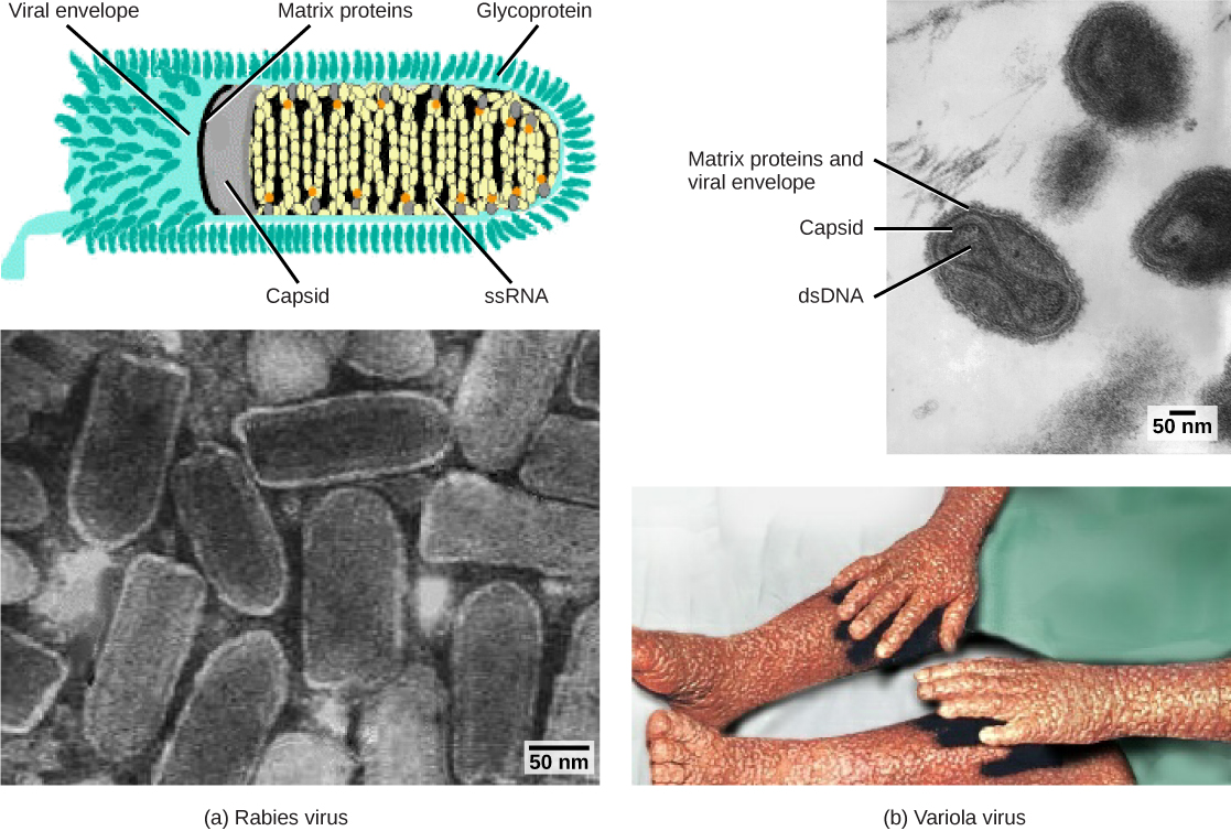
Viruses contain only a few elements by which they can be classified: the viral genome, the type of capsid, and the envelope structure for the enveloped viruses. All of these elements have been used in the past for viral classification (Table 21.1 and Figure 21.6). Viral genomes may vary in the type of genetic material (DNA or RNA) and its organization (single- or double-stranded, linear or circular, and segmented or non-segmented). In some viruses, additional proteins needed for replication are associated directly with the genome or contained within the viral capsid.
Non-segmented: genome consists of a single segment of genetic material
非分節型:ゲノムが1本の遺伝物質からなる。
Segmented: genome is divided into multiple segments
分節型:ゲノムが複数の分節に分かれる。
Parainfluenza viruses
パラインフルエンザウイルス
Influenza viruses
インフルエンザウイルス
Table 21.1表 21.1
OpenStax, Rice University; Biology 2e. (credit “rabies diagram”: modification of work by CDC; “rabies micrograph”: modification of work by Dr. Fred Murphy, CDC; credit “small pox micrograph”: modification of work by Dr. Fred Murphy, Sylvia Whitfield, CDC; credit “smallpox photo”: modification of work by CDC; scale-bar data from Matt Russell) · CC BY 4.0 · Source出典
Figure 21.6図 21.6
Viruses can be classified according to their core genetic material and capsid design. (a) Rabies virus has a single-stranded RNA (ssRNA) core and an enveloped helical capsid, whereas (b) variola virus, the causative agent of smallpox, has a double-stranded DNA (dsDNA) core and a complex capsid. Rabies transmission occurs when saliva from an infected mammal enters a wound. The virus travels through neurons in the peripheral nervous system to the central nervous system, where it impairs brain function, and then travels to other tissues. The virus can infect any mammal, and most die within weeks of infection. Smallpox is a human virus transmitted by inhalation of the variola virus, localized in the skin, mouth, and throat, which causes a characteristic rash. Before its eradication in 1979, infection resulted in a 30 to 35 percent mortality rate. (credit “rabies diagram”: modification of work by CDC; “rabies micrograph”: modification of work by Dr. Fred Murphy, CDC; credit “small pox micrograph”: modification of work by Dr. Fred Murphy, Sylvia Whitfield, CDC; credit “smallpox photo”: modification of work by CDC; scale-bar data from Matt Russell)
英語のヒント
ウイルスは、芯となる遺伝物質とカプシドの構造によって分類できる。(a)狂犬病ウイルスは一本鎖RNA(ssRNA)の芯と、エンベロープに包まれたらせん型カプシドをもつ。一方、(b)天然痘の病原体である天然痘ウイルスは、二本鎖DNA(dsDNA)の芯と複雑なカプシドをもつ。狂犬病は、感染哺乳類の唾液が傷口へ入ることで伝わる。ウイルスは末梢神経系のニューロンを通って中枢神経系へ達し、脳機能を障害した後、他の組織へ移る。どの哺乳類にも感染でき、大半は感染から数週間以内に死ぬ。天然痘は、天然痘ウイルスを吸い込むことで伝わるヒトのウイルス感染症である。皮膚、口、喉に局在し、特徴的な発疹を起こす。1979年の根絶前には、感染による死亡率は30~35%だった。(「狂犬病の図」:CDCの作品を改変。「狂犬病の顕微鏡写真」:Dr. Fred Murphy、CDCの作品を改変。「天然痘の顕微鏡写真」:Dr. Fred Murphy、Sylvia Whitfield、CDCの作品を改変。「天然痘の写真」:CDCの作品を改変。スケールバーのデータ:Matt Russell)
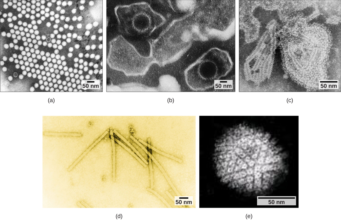
Viruses can also be classified by the design of their capsids (Table 21.2 and Figure 21.7). Capsids are classified as naked icosahedral, enveloped icosahedral, enveloped helical, naked helical, and complex. The type of genetic material (DNA or RNA) and its structure (single- or double-stranded, linear or circular, and segmented or non-segmented) are used to classify the virus core structures (Table 21.2).
Complex with many proteins; some have combinations of icosahedral and helical capsid structures
多数のタンパク質からなる複合型。一部は正二十面体型とらせん型のカプシド構造を組み合わせてもつ。
Herpesviruses, smallpox virus, hepatitis B virus, T4 bacteriophage
ヘルペスウイルス、天然痘ウイルス、B型肝炎ウイルス、T4バクテリオファージ
Table 21.2表 21.2
OpenStax, Rice University; Biology 2e. (credit a: modification of work by Dr. Fred Murphy, Sylvia Whitfield; credit b: modification of work by Liza Gross; credit c: modification of work by Dr. F. A. Murphy, CDC; credit d: modification of work by USDA ARS; credit e: modification of work by Linda Stannard, Department of Medical Microbiology, University of Cape Town, South Africa, NASA; scale-bar data from Matt Russell) · CC BY 4.0 · Source出典
Figure 21.7図 21.7
Transmission electron micrographs of various viruses show their capsid structures. The capsid of the (a) polio virus is naked icosahedral; (b) the Epstein-Barr virus capsid is enveloped icosahedral; (c) the mumps virus capsid is an enveloped helix; (d) the tobacco mosaic virus capsid is naked helical; and (e) the herpesvirus capsid is complex. (credit a: modification of work by Dr. Fred Murphy, Sylvia Whitfield; credit b: modification of work by Liza Gross; credit c: modification of work by Dr. F. A. Murphy, CDC; credit d: modification of work by USDA ARS; credit e: modification of work by Linda Stannard, Department of Medical Microbiology, University of Cape Town, South Africa, NASA; scale-bar data from Matt Russell)
各種ウイルスの透過型電子顕微鏡写真は、カプシド構造を示す。(a)ポリオウイルスは裸の正二十面体型、(b)エプスタイン・バーウイルスはエンベロープをもつ正二十面体型、(c)ムンプスウイルスはエンベロープをもつらせん型、(d)タバコモザイクウイルスは裸のらせん型、(e)ヘルペスウイルスは複合型である。(a:Dr. Fred Murphy、Sylvia Whitfieldの作品を改変。b:Liza Grossの作品を改変。c:Dr. F. A. Murphy、CDCの作品を改変。d:USDA ARSの作品を改変。e:Linda Stannard、南アフリカ・ケープタウン大学医学微生物学部門、NASAの作品を改変。スケールバーのデータ:Matt Russell)