第17章バイオテクノロジーとゲノミクス
17.1 Biotechnology17.1 バイオテクノロジー
Learning Objectives学習目標
By the end of this section, you will be able to do the following:
この節を終えると、次のことができるようになる。
- Describe gel electrophoresisゲル電気泳動を説明する。
- Explain molecular and reproductive cloning分子クローニングと生殖クローニングを説明する。
- Describe biotechnology uses in medicine and agriculture医療と農業でのバイオテクノロジーの利用を説明する。
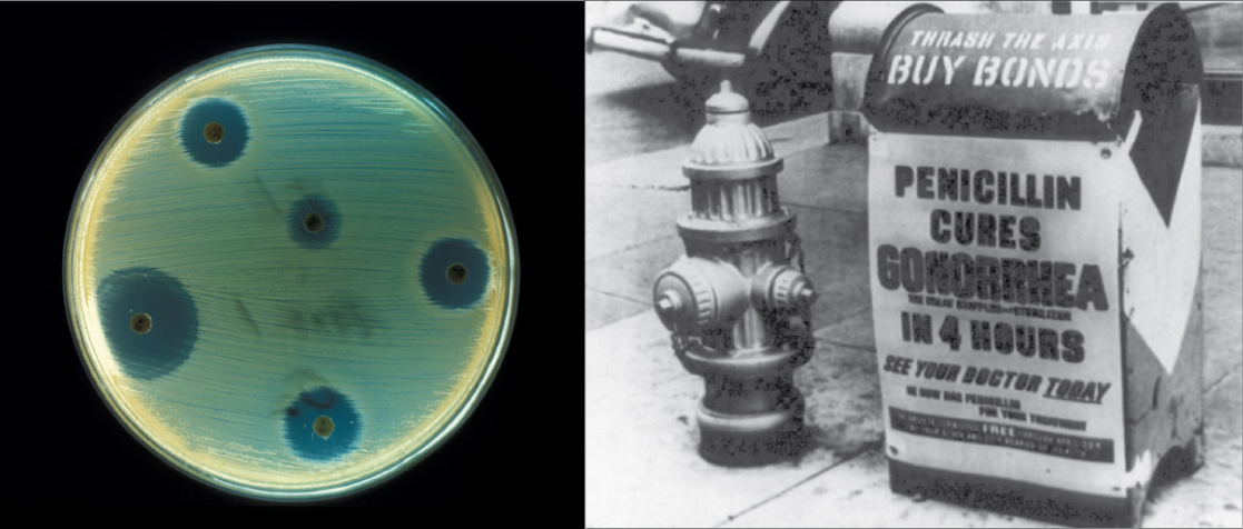
Biotechnology is the use of biological agents for technological advancement. Biotechnology was used for breeding livestock and crops long before people understood the scientific basis of these techniques. Since the discovery of the structure of DNA in 1953, the biotechnology field has grown rapidly through both academic research and private companies. The primary applications of this technology are in medicine (vaccine and antibiotic production) and agriculture (crop genetic modification in order to increase yields). Biotechnology also has many industrial applications, such as fermentation, treating oil spills, and producing biofuels (Figure 17.2).
英語のヒント
バイオテクノロジーとは、技術の進歩のために生物学的な作用をもつものを利用することである。人々がその技術の科学的な基礎を理解するはるか以前から、バイオテクノロジーは家畜や作物の品種改良に使われていた。1953年のDNA構造の発見以来、この分野は学術研究と民間企業の両方を通じて急速に発展してきた。主な応用先は、医療(ワクチンや抗生物質の生産)と農業(収量を増やすための作物の遺伝子改変)である。発酵、流出した石油の処理、バイオ燃料の生産など、工業上の応用も数多くある(図17.2)。
OpenStax, Rice University; Biology 2e. (credit "advertisement": modification of work by NIH; credit "test plate": modification of work by Don Stalons/CDC; scale-bar data from Matt Russell) · CC BY 4.0 · Source出典
Figure 17.2図 17.2
Fungi, bacteria, and other organisms that have antimicrobial properties produce antibiotics. The first antibiotic discovered was penicillin. Pharmaceutical companies now commercially produce and test antibiotics for their potential to inhibit bacterial growth. (credit "advertisement": modification of work by NIH; credit "test plate": modification of work by Don Stalons/CDC; scale-bar data from Matt Russell)
英語のヒント
抗菌作用をもつ菌類、細菌、その他の生物は、抗生物質を産生する。最初に発見された抗生物質はペニシリンだった。現在、製薬会社は抗生物質を商業的に生産し、細菌の増殖を阻害する可能性を試験している。(「広告」のクレジット:NIHの作品を改変。「試験用プレート」のクレジット:Don Stalons/CDCの作品を改変。スケールバーのデータ:Matt Russell)
Basic Techniques to Manipulate Genetic Material (DNA and RNA)遺伝物質(DNAとRNA)を操作する基本技術
To understand the basic techniques used to work with nucleic acids, remember that nucleic acids are macromolecules made of nucleotides (a sugar, a phosphate, and a nitrogenous base) linked by phosphodiester bonds. The phosphate groups on these molecules each have a net negative charge. An entire set of DNA molecules in the nucleus is called the genome. DNA has two complementary strands linked by hydrogen bonds between the paired bases. Exposure to high temperatures (DNA denaturation) can separate the two strands and cooling can reanneal them. The DNA polymerase enzyme can replicate the DNA. Unlike DNA, which is located in the eukaryotic cells' nucleus, RNA molecules leave the nucleus. The most common type of RNA that researchers analyze is the messenger RNA (mRNA) because it represents the protein-coding genes that are actively expressed. However, RNA molecules present some other challenges to analysis, as they are often less stable than DNA.
英語のヒント
核酸を扱う基本技術を理解するために、核酸はヌクレオチド(糖、リン酸、窒素を含む塩基)がホスホジエステル結合でつながった高分子であることを思い出そう。これらの分子のリン酸基は、それぞれ正味の負電荷をもつ。核内のDNA分子の一式をゲノムと呼ぶ。DNAには相補的な2本の鎖があり、対をなす塩基間の水素結合で結ばれている。高温にさらすこと(DNAの変性)で2本の鎖を分離でき、冷やすと再び結合させることができる。DNAポリメラーゼという酵素は、DNAを複製できる。真核細胞の核内にあるDNAとは異なり、RNA分子は核から出る。研究者が最もよく分析するRNAはメッセンジャーRNA(mRNA)である。mRNAは、活発に発現しているタンパク質コード遺伝子を反映するためである。しかしRNA分子は、DNAより不安定であることが多く、分析にはさらに別の難しさがある。
DNA and RNA ExtractionDNAとRNAの抽出
To study or manipulate nucleic acids, one must first isolate or extract the DNA or RNA from the cells. Researchers use various techniques to extract different types of DNA (Figure 17.3). Most nucleic acid extraction techniques involve steps to break open the cell and use enzymatic reactions to destroy all macromolecules that are not desired (such as unwanted molecule degradation and separation from the DNA sample). A lysis buffer (a solution which is mostly a detergent) breaks cells. Note that lysis means "to split". These enzymes break apart lipid molecules in the cell membranes and nuclear membranes. Enzymes such as proteases and ribonucleases (RNases) break down proteins and RNA, respectively. Using alcohol precipitates the DNA. Human genomic DNA is usually visible as a gelatinous, white mass. One can store the DNA samples frozen at –80°C for several years.
英語のヒント
核酸を研究したり操作したりするには、まず細胞からDNAやRNAを分離、すなわち抽出しなければならない。研究者は、異なる種類のDNAを抽出するためにさまざまな技術を使う(図17.3)。核酸抽出の多くの方法には、細胞を壊して開き、酵素反応を用いて不要な高分子をすべて破壊する段階が含まれる(不要な分子の分解やDNA試料からの分離など)。溶解バッファー(主として界面活性剤からなる溶液)は細胞を壊す。lysisは「分裂させる」という意味であることに注意しよう。これらの酵素は、細胞膜や核膜の脂質分子を分解する。プロテアーゼやリボヌクレアーゼ(RNase)などの酵素は、それぞれタンパク質とRNAを分解する。アルコールを用いると、DNAが沈殿する。ヒトのゲノムDNAは通常、ゼラチン状の白い塊として見える。DNA試料は、−80°Cで凍結させて数年間保存できる。
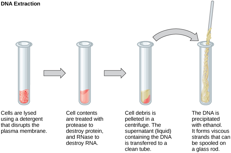


Figure 17.3図 17.3
This diagram shows the basic method of DNA extraction.
DNA抽出の基本的な方法を示した図。
Scientists perform RNA analysis to study gene expression patterns in cells. RNA is naturally very unstable because RNAses are commonly present in nature and very difficult to inactivate. Similar to DNA, RNA extraction involves using various buffers and enzymes to inactivate macromolecules and preserve the RNA.
英語のヒント
科学者は、細胞の遺伝子発現パターンを調べるためにRNAを分析する。RNaseは自然界に広く存在し、不活性化するのが非常に難しいため、RNAはもともときわめて不安定である。DNAと同様に、RNAの抽出でも、さまざまな緩衝液や酵素を用いて高分子を不活性化し、RNAを保存する。
Gel Electrophoresisゲル電気泳動
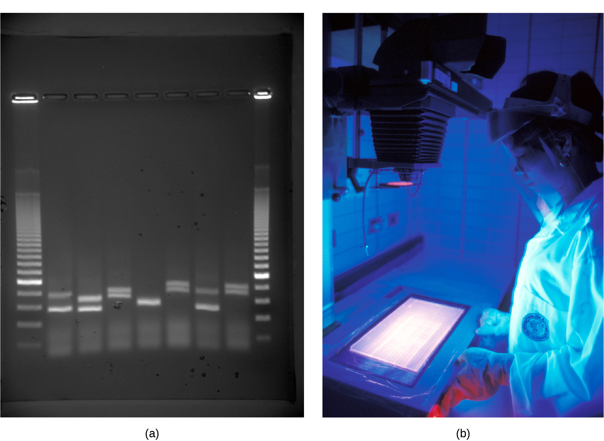
Because nucleic acids are negatively charged ions at neutral or basic pH in an aqueous environment, an electric field can mobilize them. Gel electrophoresis is a technique that scientists use to separate molecules on the basis of size, using this charge. One can separate the nucleic acids as whole chromosomes or fragments. The nucleic acids load into a slot near the semisolid, porous gel matrix's negative electrode, and pulled toward the positive electrode at the gel's opposite end. Smaller molecules move through the gel's pores faster than larger molecules. This difference in the migration rate separates the fragments on the basis of size. There are molecular weight standard samples that researchers can run alongside the molecules to provide a size comparison. We can observe nucleic acids in a gel matrix using various fluorescent or colored dyes. Distinct nucleic acid fragments appear as bands at specific distances from the gel's top (the negative electrode end) on the basis of their size (Figure 17.4). A mixture of genomic DNA fragments of varying sizes appear as a long smear; whereas, uncut genomic DNA is usually too large to run through the gel and forms a single large band at the gel's top.
英語のヒント
核酸は水中で、中性または塩基性のpHでは負に帯電したイオンになるため、電場によって移動させることができる。ゲル電気泳動は、この電荷を利用して、分子を大きさに基づいて分離する技術である。核酸は、染色体全体の状態でも、断片の状態でも分離できる。核酸を、半固体で多孔質のゲル基質の負極近くにある溝へ入れると、ゲルの反対側の端にある正極へ引かれる。小さな分子は、大きな分子よりも速くゲルの孔を通り抜ける。この移動速度の違いによって、断片が大きさに応じて分離される。大きさを比較するために、試料の分子と並べて泳動できる分子量標準試料がある。さまざまな蛍光色素や着色色素を用いると、ゲル基質中の核酸を観察できる。それぞれの核酸断片は、その大きさに応じて、ゲルの上端(負極側)から特定の距離のところに帯として現れる(図17.4)。大きさの異なるゲノムDNA断片の混合物は、長いにじみ状の像となる。一方、切断していないゲノムDNAは通常、大きすぎてゲル中を移動できず、ゲルの上端に一つの大きな帯を作る。
OpenStax, Rice University; Biology 2e. (credit: a: James Jacob, Tompkins Cortland Community College b: International Rice Research Institute) · CC BY 4.0 · Source出典

Figure 17.4図 17.4
a) Shown are DNA fragments from seven samples run on a gel, stained with a fluorescent dye, and viewed under UV light; and b) a researcher from International Rice Research Institute, reviewing DNA profiles using UV light. (credit: a: James Jacob, Tompkins Cortland Community College b: International Rice Research Institute)
a)7試料のDNA断片をゲルで泳動し、蛍光色素で染色して紫外線下で観察したもの。b)紫外線を用いてDNAのパターンを確認する、国際稲研究所の研究者。(クレジット:a:James Jacob、Tompkins Cortland Community College。b:International Rice Research Institute)