第40章循環器系
40.4 Blood Flow and Blood Pressure Regulation40.4 血流と血圧の調節
Learning Objectives学習目標
By the end of this section, you will be able to do the following:
この節を終えると、次のことができるようになる。
- Describe the system of blood flow through the body体内を流れる血液の仕組みを説明する。
- Describe how blood pressure is regulated血圧がどのように調節されるかを説明する。
Blood pressure (BP) is the pressure exerted by blood on the walls of a blood vessel that helps to push blood through the body. Systolic blood pressure measures the amount of pressure that blood exerts on vessels while the heart is beating. The optimal systolic blood pressure is 120 mm Hg. Diastolic blood pressure measures the pressure in the vessels between heartbeats. The optimal diastolic blood pressure is 80 mm Hg. Many factors can affect blood pressure, such as hormones, stress, exercise, eating, sitting, and standing. Blood flow through the body is regulated by the size of blood vessels, by the action of smooth muscle, by one-way valves, and by the fluid pressure of the blood itself.
英語のヒント
血圧(BP)は、血液が血管壁に及ぼす圧力であり、血液を体内へ押し流すのを助ける。収縮期血圧は、心臓が拍動しているときに血液が血管に及ぼす圧力を表し、最適値は120mmHgである。拡張期血圧は、拍動と拍動の間の血管内圧を表し、最適値は80mmHgである。ホルモン、ストレス、運動、食事、座位、立位など、多くの要因が血圧に影響しうる。体内の血流は、血管の太さ、平滑筋の作用、一方向に開く弁、血液自体の流体圧によって調節される。
How Blood Flows Through the Body体内を流れる血液
Blood is pushed through the body by the action of the pumping heart. With each rhythmic pump, blood is pushed under high pressure and velocity away from the heart, initially along the main artery, the aorta. In the aorta, the blood travels at 30 cm/sec. As blood moves into the arteries, arterioles, and ultimately to the capillary beds, the rate of movement slows dramatically to about 0.026 cm/sec, one-thousand times slower than the rate of movement in the aorta. While the diameter of each individual arteriole and capillary is far narrower than the diameter of the aorta, and according to the law of continuity, fluid should travel faster through a narrower diameter tube, the rate is actually slower due to the overall diameter of all the combined capillaries being far greater than the diameter of the individual aorta.
英語のヒント
血液は、心臓のポンプ作用によって体内へ押し流される。規則的な拍動のたびに、高い圧力と速度で心臓から送り出され、まず主要な動脈である大動脈を進む。大動脈での流速は30cm/秒である。血液が動脈、細動脈、最終的には毛細血管床へ進むにつれ、流速は大きく下がり、大動脈の約1000分の1の0.026cm/秒になる。個々の細動脈や毛細血管は大動脈よりはるかに細く、連続の法則に従えば、細い管では液体は速く流れるはずである。それでも実際には遅くなるのは、すべての毛細血管を合わせた全体の直径が、1本の大動脈の直径よりはるかに大きいためである。
The slow rate of travel through the capillary beds, which reach almost every cell in the body, assists with gas and nutrient exchange and also promotes the diffusion of fluid into the interstitial space. After the blood has passed through the capillary beds to the venules, veins, and finally to the main venae cavae, the rate of flow increases again but is still much slower than the initial rate in the aorta. Blood primarily moves in the veins by the rhythmic movement of smooth muscle in the vessel wall and by the action of the skeletal muscle as the body moves. Because most veins must move blood against the pull of gravity, blood is prevented from flowing backward in the veins by one-way valves. Because skeletal muscle contraction aids in venous blood flow, it is important to get up and move frequently after long periods of sitting so that blood will not pool in the extremities.
英語のヒント
毛細血管床は体のほぼすべての細胞へ届き、そこを血液がゆっくり流れることで、気体や栄養素の交換が助けられ、間質腔への液体の拡散も促される。血液が毛細血管床から細静脈、静脈、最後に主要な大静脈へ進むと流速は再び上がるが、大動脈での最初の速度よりはかなり遅い。静脈内の血液は、主に血管壁の平滑筋の規則的な動きと、体が動くときの骨格筋の作用で移動する。多くの静脈は重力に逆らって血液を運ぶ必要があるため、一方向に開く弁が逆流を防ぐ。骨格筋の収縮は静脈血の流れを助ける。そのため、長く座った後にはこまめに立って動き、四肢に血液がたまらないようにすることが重要である。
Blood flow through the capillary beds is regulated depending on the body’s needs and is directed by nerve and hormone signals. For example, after a large meal, most of the blood is diverted to the stomach by vasodilation of vessels of the digestive system and vasoconstriction of other vessels. During exercise, blood is diverted to the skeletal muscles through vasodilation while blood to the digestive system would be lessened through vasoconstriction. The blood entering some capillary beds is controlled by small muscles, called precapillary sphincters, illustrated in Figure 40.17. If the sphincters are open, the blood will flow into the associated branches of the capillary blood. If all of the sphincters are closed, then the blood will flow directly from the arteriole to the venule through the thoroughfare channel (see Figure 40.17). These muscles allow the body to precisely control when capillary beds receive blood flow. At any given moment only about 5–10% of our capillary beds actually have blood flowing through them.
英語のヒント
毛細血管床の血流は、体の必要に応じて、神経やホルモンの信号によって調節される。たとえば大量に食事をすると、消化器系の血管が拡張し、ほかの血管が収縮して、多くの血液が胃へ向けられる。運動中には、血管拡張によって骨格筋へ血液が向かい、消化器系への血液は血管収縮によって減る。一部の毛細血管床へ入る血液は、図40.17に示す前毛細血管括約筋という小さな筋で制御される。括約筋が開けば、対応する毛細血管の枝へ血液が流れる。すべて閉じると、血液は直通路を通り、細動脈から細静脈へ直接流れる(図40.17)。これらの筋によって、毛細血管床に血液を流す時期を正確に制御できる。ある瞬間に実際に血液が流れているのは、毛細血管床の約5〜10%だけである。
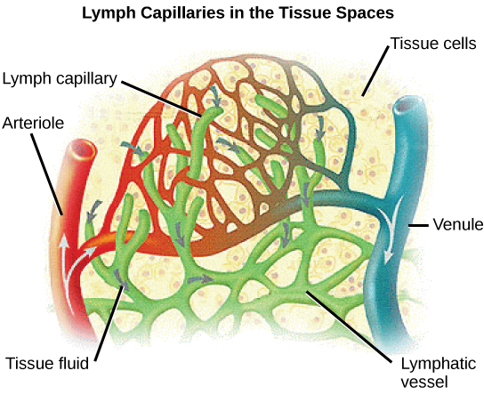
Proteins and other large solutes cannot leave the capillaries. The loss of the watery plasma creates a hyperosmotic solution within the capillaries, especially near the venules. This causes about 85% of the plasma that leaves the capillaries to eventually diffuse back into the capillaries near the venules. The remaining 15% of blood plasma drains out from the interstitial fluid into nearby lymphatic vessels (Figure 40.18). The fluid in the lymph is similar in composition to the interstitial fluid. The lymph fluid passes through lymph nodes before it returns to the heart via the superior vena cava. Lymph nodes are specialized organs that filter the lymph by percolation through a maze of connective tissue filled with white blood cells. The white blood cells remove infectious agents, such as bacteria and viruses, to clean the lymph before it returns to the bloodstream. After it is cleaned, the lymph returns to the heart by the action of smooth muscle pumping, skeletal muscle action, and one-way valves joining the returning blood near the junction of the venae cavae entering the right atrium of the heart.
英語のヒント
タンパク質などの大きな溶質は、毛細血管から出ることができない。水分の多い血漿が失われると、毛細血管内、特に細静脈に近い部分の溶液が高浸透圧になる。そのため、毛細血管を出た血漿の約85%は、最終的に細静脈の近くで毛細血管内へ拡散して戻る。残りの15%は、間質液から近くのリンパ管へ流れ込む(図40.18)。リンパ液の組成は間質液に似ている。リンパ液はリンパ節を通過し、その後、上大静脈を経て心臓へ戻る。リンパ節は、白血球を多く含む迷路状の結合組織にリンパを通し、ろ過する特殊な器官である。白血球は細菌やウイルスなどの感染因子を除去し、血流へ戻る前にリンパを浄化する。浄化されたリンパは、平滑筋のポンプ作用、骨格筋の作用、一方向に開く弁によって心臓へ戻る。右心房に入る大静脈の合流部付近で、戻ってくる血液に合流する。
OpenStax, Rice University; Biology 2e. (credit: modification of work by NCI, NIH) · CC BY 4.0 · Source出典

Figure 40.18図 40.18
Fluid from the capillaries moves into the interstitial space and lymph capillaries by diffusion down a pressure gradient and also by osmosis. Out of 7,200 liters of fluid pumped by the average heart in a day, over 1,500 liters is filtered. (credit: modification of work by NCI, NIH)
毛細血管の液体は、圧力勾配に従う拡散と浸透によって、間質腔やリンパ毛細管へ移動する。平均的な心臓が1日に送る7200リットルの液体のうち、1500リットル以上がろ過される。(出典:NCI、NIHの作品を改変)