第40章循環器系
White Blood Cells白血球
White blood cells, also called leukocytes (leuko = white), make up approximately one percent by volume of the cells in blood. The role of white blood cells is very different than that of red blood cells: they are primarily involved in the immune response to identify and target pathogens, such as invading bacteria, viruses, and other foreign organisms. White blood cells are formed continually; some only live for hours or days, but some live for years.
英語のヒント
白血球は英語でleukocytesとも呼ばれる(leukoは「白」)。血液中の細胞の体積の約1%を占める。その役割は赤血球とは大きく異なり、主に免疫応答に関わる。侵入した細菌、ウイルスなどの外来生物を病原体として認識し、攻撃の標的とする。白血球は絶えずつくられている。寿命が数時間や数日のものもあれば、数年生きるものもある。
The morphology of white blood cells differs significantly from red blood cells. They have nuclei and do not contain hemoglobin. The different types of white blood cells are identified by their microscopic appearance after histologic staining, and each has a different specialized function. The two main groups, both illustrated in Figure 40.7 are the granulocytes, which include the neutrophils, eosinophils, and basophils, and the agranulocytes, which include the monocytes and lymphocytes.
英語のヒント
白血球の形態は、赤血球とは大きく異なる。白血球には核があり、ヘモグロビンはない。種類の違いは、組織学的な染色後の顕微鏡像によって区別でき、それぞれが異なる専門の機能をもつ。図40.7に示す2つの主要な群は、好中球、好酸球、好塩基球を含む顆粒球と、単球、リンパ球を含む無顆粒球である。
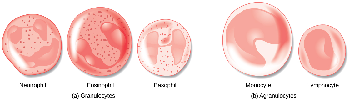
Figure 40.7図 40.7
(a) Granulocytes—including neutrophils, eosinophils and basophils—are characterized by a lobed nucleus and granular inclusions in the cytoplasm. Granulocytes are typically first-responders during injury or infection. (b) Agranulocytes include lymphocytes and monocytes. Lymphocytes, including B and T cells, are responsible for adaptive immune response. Monocytes differentiate into macrophages and dendritic cells, which in turn respond to infection or injury.
(a)好中球、好酸球、好塩基球を含む顆粒球は、分葉した核と、細胞質内の顆粒状の構造を特徴とする。一般に、損傷や感染に最初に応答する。(b)無顆粒球にはリンパ球と単球が含まれる。B細胞とT細胞を含むリンパ球は、適応免疫応答を担う。単球はマクロファージや樹状細胞へ分化し、これらも感染や損傷に応答する。
Granulocytes contain granules in their cytoplasm; the agranulocytes are so named because of the lack of granules in their cytoplasm. Some leukocytes become macrophages that either stay at the same site or move through the bloodstream and gather at sites of infection or inflammation where they are attracted by chemical signals from foreign particles and damaged cells. Lymphocytes are the primary cells of the immune system and include B cells, T cells, and natural killer cells. B cells destroy bacteria and inactivate their toxins. They also produce antibodies. T cells attack viruses, fungi, some bacteria, transplanted cells, and cancer cells. T cells attack viruses by releasing toxins that kill the viruses. Natural killer cells attack a variety of infectious microbes and certain tumor cells.
英語のヒント
顆粒球には細胞質内に顆粒がある。無顆粒球という名前は、細胞質内に顆粒がないことに由来する。一部の白血球はマクロファージになる。マクロファージは同じ場所にとどまるか、血流を移動して感染部位や炎症部位に集まる。外来粒子や損傷細胞からの化学的な信号に引き寄せられるためである。リンパ球は免疫系の中心的な細胞で、B細胞、T細胞、ナチュラルキラー細胞を含む。B細胞は細菌を破壊し、その毒素を不活化する。また、抗体もつくる。T細胞はウイルス、真菌、一部の細菌、移植細胞、がん細胞を攻撃する。T細胞は、ウイルスを殺す毒素を放出してウイルスを攻撃する。ナチュラルキラー細胞は、さまざまな感染性微生物や、特定の腫瘍細胞を攻撃する。
One reason that HIV poses significant management challenges is because the virus directly targets T cells by gaining entry through a receptor. Once inside the cell, HIV then multiplies using the T cell’s own genetic machinery. After the HIV virus replicates, it is transmitted directly from the infected T cell to macrophages. The presence of HIV can remain unrecognized for an extensive period of time before full disease symptoms develop.
英語のヒント
HIVの管理が非常に難しい理由の1つは、ウイルスが受容体を介して入り込み、T細胞を直接の標的にすることである。いったん細胞内に入ると、HIVはT細胞自身の遺伝的な仕組みを使って増殖する。複製後のHIVは、感染したT細胞からマクロファージへ直接伝わる。病気の症状が十分に現れるまで、長い間、HIVの存在が認識されないこともある。
Platelets and Coagulation Factors血小板と凝固因子
Blood must clot to heal wounds and prevent excess blood loss. Small cell fragments called platelets (thrombocytes) are attracted to the wound site where they adhere by extending many projections and releasing their contents. These contents activate other platelets and also interact with other coagulation factors, which convert fibrinogen, a water-soluble protein present in blood serum into fibrin (a non-water soluble protein), causing the blood to clot. Many of the clotting factors require vitamin K to work, and vitamin K deficiency can lead to problems with blood clotting. Many platelets converge and stick together at the wound site forming a platelet plug (also called a fibrin clot), as illustrated in Figure 40.8b. The plug or clot lasts for a number of days and stops the loss of blood. Platelets are formed from the disintegration of larger cells called megakaryocytes, like that shown in Figure 40.8a. For each megakaryocyte, 2000–3000 platelets are formed with 150,000 to 400,000 platelets present in each cubic millimeter of blood. Each platelet is disc shaped and 2–4 μm in diameter. They contain many small vesicles but do not contain a nucleus.
英語のヒント
傷を治し、過剰な出血を防ぐために、血液は凝固しなければならない。血小板という小さな細胞片が傷の部位へ引き寄せられ、多数の突起を伸ばし、内容物を放出しながら付着する。放出された内容物は、ほかの血小板を活性化し、ほかの凝固因子とも相互作用する。凝固因子は、血清中にある水溶性タンパク質のフィブリノゲンを、水に溶けないフィブリンへ変え、血液を凝固させる。多くの凝固因子は働くためにビタミンKを必要とするため、その欠乏は血液凝固の障害につながりうる。図40.8bのように、多数の血小板が傷の部位に集まって互いに付着し、血小板栓(フィブリン血餅とも呼ばれる)を形成する。この栓、すなわち血餅は数日間残り、出血を止める。血小板は、図40.8aのような、巨核球という大きな細胞が分断されてつくられる。巨核球1個から2000〜3000個の血小板が生じ、血液1立方ミリメートルあたり15万〜40万個が存在する。各血小板は直径2〜4µmの円盤状で、多数の小胞を含むが、核はない。
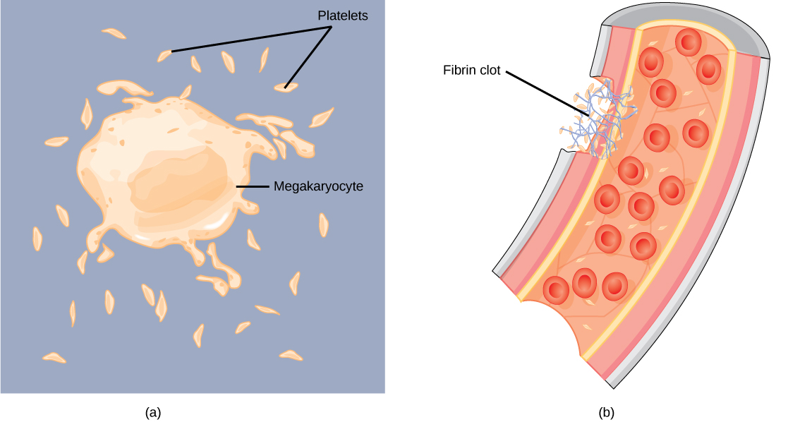


Figure 40.8図 40.8
(a) Platelets are formed from large cells called megakaryocytes. The megakaryocyte breaks up into thousands of fragments that become platelets. (b) Platelets are required for clotting of the blood. The platelets collect at a wound site in conjunction with other clotting factors, such as fibrinogen, to form a fibrin clot that prevents blood loss and allows the wound to heal.
(a)血小板は、巨核球という大きな細胞からつくられる。巨核球が数千個の断片に分かれ、それらが血小板となる。(b)血小板は血液凝固に必要である。傷の部位に集まり、フィブリノゲンなどの凝固因子とともにフィブリン血餅を形成する。これが出血を防ぎ、傷の治癒を可能にする。