第40章循環器系
The Cardiac Cycle心周期
The main purpose of the heart is to pump blood through the body; it does so in a repeating sequence called the cardiac cycle. The cardiac cycle is the coordination of the filling and emptying of the heart of blood by electrical signals that cause the heart muscles to contract and relax. The human heart beats over 100,000 times per day. In each cardiac cycle, the heart contracts (systole), pushing out the blood and pumping it through the body; this is followed by a relaxation phase (diastole), where the heart fills with blood, as illustrated in Figure 40.12. The atria contract at the same time, forcing blood through the atrioventricular valves into the ventricles. Closing of the atrioventricular valves produces a monosyllabic “lup” sound. Following a brief delay, the ventricles contract at the same time forcing blood through the semilunar valves into the aorta and the artery transporting blood to the lungs (via the pulmonary artery). Closing of the semilunar valves produces a monosyllabic “dup” sound.
英語のヒント
心臓の主な役割は、体内へ血液を送り出すことであり、心周期という一連の動きを繰り返して行う。心周期では、電気信号が心筋を収縮・弛緩させ、心臓が血液で満たされる動きと、血液を送り出す動きが協調して進む。ヒトの心臓は1日に10万回以上拍動する。各心周期では、心臓が収縮し(収縮期)、血液を押し出して全身へ送る。その後、図40.12のように、心臓が血液で満たされる弛緩の時期(拡張期)が続く。左右の心房は同時に収縮し、血液を房室弁から心室へ押し込む。房室弁が閉じると、「ラッ」という1音節の音がする。少し遅れて左右の心室が同時に収縮し、血液を半月弁から大動脈と肺へ向かう動脈(肺動脈)へ押し出す。半月弁が閉じると、「ダッ」という1音節の音がする。
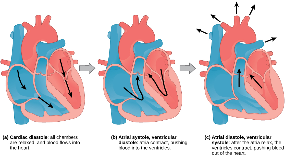
Figure 40.12図 40.12
During (a) cardiac diastole, the heart muscle is relaxed and blood flows into the heart. During (b) atrial systole, the atria contract, pushing blood into the ventricles. During (c) atrial diastole, the ventricles contract, forcing blood out of the heart.
(a)心臓の拡張期には心筋が弛緩し、血液が心臓へ流れ込む。(b)心房の収縮期には心房が収縮し、心室へ血液を押し込む。(c)心房の拡張期には心室が収縮し、心臓の外へ血液を押し出す。
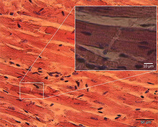
The pumping of the heart is a function of the cardiac muscle cells, or cardiomyocytes, that make up the heart muscle. Cardiomyocytes, shown in Figure 40.13, are distinctive muscle cells that are striated like skeletal muscle but pump rhythmically and involuntarily like smooth muscle; they are connected by intercalated disks exclusive to cardiac muscle. They are self-stimulated for a period of time and isolated cardiomyocytes will beat if given the correct balance of nutrients and electrolytes.
英語のヒント
心臓のポンプ作用は、心筋を構成する心筋細胞の働きである。図40.13の心筋細胞は、骨格筋のように横紋をもつ一方、平滑筋のように不随意に、リズミカルに収縮する独特の筋細胞である。細胞どうしは、心筋に特有の介在板でつながる。心筋細胞は一定時間、自ら刺激を生み出す。単離した心筋細胞も、栄養素と電解質を適切なバランスで与えると拍動する。
OpenStax, Rice University; Biology 2e. (credit: modification of work by Dr. S. Girod, Anton Becker; scale-bar data from Matt Russell) · CC BY 4.0 · Source出典
Figure 40.13図 40.13
Cardiomyocytes are striated muscle cells found in cardiac tissue. (credit: modification of work by Dr. S. Girod, Anton Becker; scale-bar data from Matt Russell)
心筋細胞は、心臓の組織にある横紋をもつ筋細胞である。(出典:Dr. S. Girod、Anton Beckerの作品を改変。スケールバーのデータ:Matt Russell)
The autonomous beating of cardiac muscle cells is regulated by the heart’s internal pacemaker that uses electrical signals to time the beating of the heart. The electrical signals and mechanical actions, illustrated in Figure 40.14, are intimately intertwined. The internal pacemaker starts at the sinoatrial (SA) node, which is located near the wall of the right atrium. Electrical charges spontaneously pulse from the SA node causing the two atria to contract in unison. The pulse reaches a second node, called the atrioventricular (AV) node, between the right atrium and right ventricle where it pauses for approximately 0.1 second before spreading to the walls of the ventricles. From the AV node, the electrical impulse enters the bundle of His, then to the left and right bundle branches extending through the interventricular septum. Finally, the Purkinje fibers conduct the impulse from the apex of the heart up the ventricular myocardium, and then the ventricles contract. This pause allows the atria to empty completely into the ventricles before the ventricles pump out the blood. The electrical impulses in the heart produce electrical currents that flow through the body and can be measured on the skin using electrodes. This information can be observed as an electrocardiogram (ECG)—a recording of the electrical impulses of the cardiac muscle.
英語のヒント
心筋細胞の自律的な拍動は、電気信号で心拍のタイミングを調節する、心臓内部のペースメーカーによって制御される。図40.14に示す電気信号と機械的な動きは、密接に結び付いている。内部のペースメーカーは、右心房の壁の近くにある洞房結節(SA結節)から始まる。SA結節から電気的な刺激が自発的に発生し、左右の心房を同時に収縮させる。刺激は右心房と右心室の間にある第2の結節、房室結節(AV結節)へ達し、約0.1秒遅れてから心室壁へ広がる。AV結節から出た電気的興奮はヒス束に入り、次いで心室中隔を通って伸びる左脚と右脚へ伝わる。最後にプルキンエ線維が、心尖から心室の心筋を上方へ向かって興奮を伝え、心室が収縮する。この遅れにより、心室が血液を送り出す前に、心房内の血液が十分に心室へ流れ込む。心臓の電気的興奮は体内を流れる電流を生み、皮膚上の電極で測定できる。この情報は、心筋の電気的興奮を記録する心電図(ECG)として観察できる。
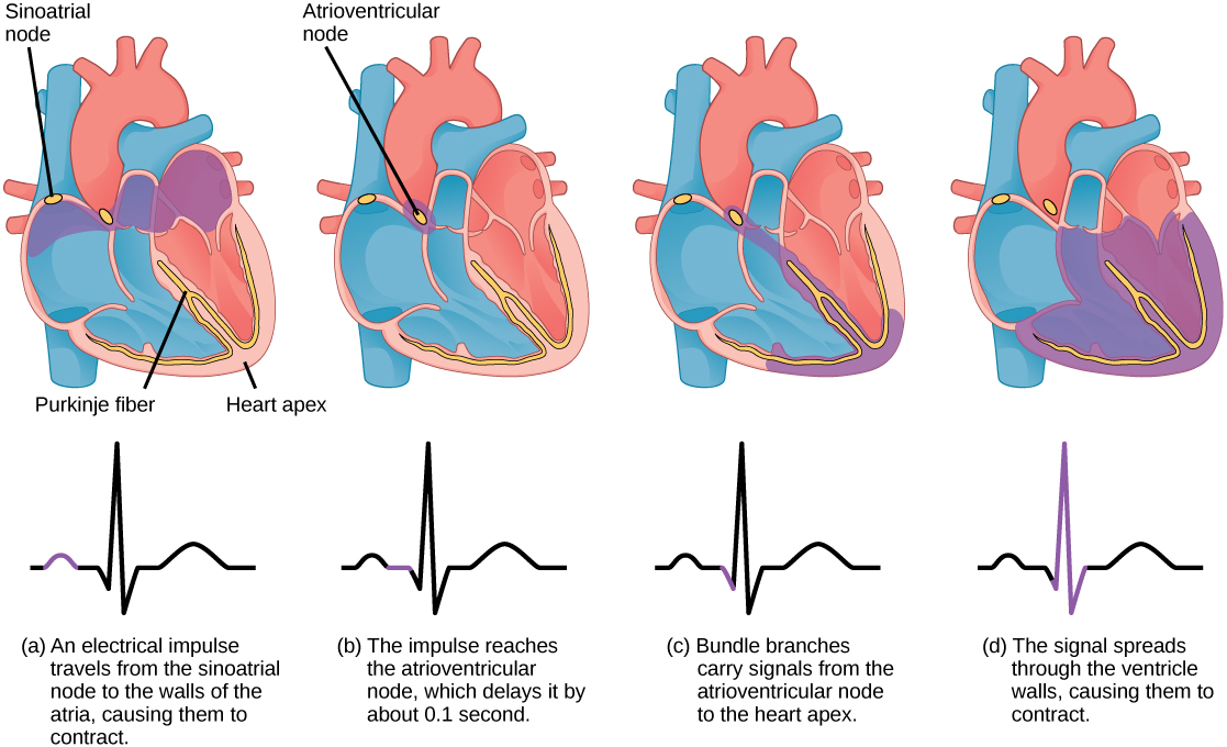


Figure 40.14図 40.14
The beating of the heart is regulated by an electrical impulse that causes the characteristic reading of an ECG. The signal is initiated at the sinoatrial node. The signal then (a) spreads to the atria, causing them to contract. The signal is (b) delayed at the atrioventricular node before it is passed on to the (c) heart apex. The delay allows the atria to relax before the (d) ventricles contract. The final part of the ECG cycle prepares the heart for the next beat.
心拍は電気的興奮によって調節され、それが心電図の特徴的な波形を生む。信号は洞房結節で始まり、(a)心房へ広がって収縮させる。(b)房室結節でいったん遅れた後、(c)心尖へ伝わる。この遅れにより、(d)心室が収縮する前に心房が弛緩できる。心電図の周期の最後の部分では、次の拍動に備える。
Arteries, Veins, and Capillaries動脈・静脈・毛細血管
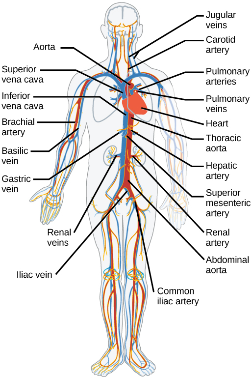
The blood from the heart is carried through the body by a complex network of blood vessels (Figure 40.15). Arteries take blood away from the heart. The main artery is the aorta that branches into major arteries that take blood to different limbs and organs. These major arteries include the carotid artery that takes blood to the brain, the brachial arteries that take blood to the arms, and the thoracic artery that takes blood to the thorax and then into the hepatic, renal, and gastric arteries for the liver, kidney, and stomach, respectively. The iliac artery takes blood to the lower limbs. The major arteries diverge into minor arteries, and then smaller vessels called arterioles, to reach more deeply into the muscles and organs of the body.
英語のヒント
心臓から出た血液は、複雑な血管網によって全身へ運ばれる(図40.15)。動脈は、心臓から離れる方向へ血液を運ぶ。主な動脈は大動脈であり、四肢や各器官へ血液を送る主要な動脈に枝分かれする。主要な動脈には、脳へ血液を送る頸動脈、腕へ送る上腕動脈、胸部へ送る胸部の動脈がある。胸部の動脈からは、肝臓、腎臓、胃へ向かう肝動脈、腎動脈、胃動脈へ血液が流れる。腸骨動脈は下肢へ血液を送る。主要な動脈は細い動脈へ、さらに細動脈という小さな血管へ分かれ、筋や器官のより深いところへ届く。
OpenStax, Rice University; Biology 2e. (credit: modification of work by Mariana Ruiz Villareal) · CC BY 4.0 · Source出典

Figure 40.15図 40.15
The major human arteries and veins are shown. (credit: modification of work by Mariana Ruiz Villareal)
ヒトの主な動脈と静脈を示す。(出典:Mariana Ruiz Villarealの作品を改変)
Arterioles diverge into capillary beds. Capillary beds contain a large number (10 to 100) of capillaries that branch among the cells and tissues of the body. Capillaries are narrow-diameter tubes that can fit red blood cells through in single file and are the sites for the exchange of nutrients, waste, and oxygen with tissues at the cellular level. Fluid also crosses into the interstitial space from the capillaries. The capillaries converge again into venules that connect to minor veins that finally connect to major veins that take blood high in carbon dioxide back to the heart. Veins are blood vessels that bring blood back to the heart. The major veins drain blood from the same organs and limbs that the major arteries supply. Fluid is also brought back to the heart via the lymphatic system.
英語のヒント
細動脈は枝分かれして毛細血管床になる。毛細血管床には、体の細胞や組織の間で分岐する、多数の毛細血管(10〜100本)が含まれる。毛細血管は直径が小さく、赤血球が1列で通るほど細い管である。細胞レベルで、組織と栄養素、老廃物、酸素を交換する場所となる。液体も毛細血管から間質腔へ移動する。毛細血管は再び集まって細静脈となり、細い静脈から主要な静脈へつながる。これらが二酸化炭素の多い血液を心臓へ戻す。静脈とは、心臓へ血液を戻す血管である。主要な静脈は、主要な動脈が血液を供給するのと同じ器官や四肢から血液を集める。液体はリンパ系を介しても心臓へ戻される。
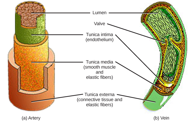
The structure of the different types of blood vessels reflects their function or layers. There are three distinct layers, or tunics, that form the walls of blood vessels (Figure 40.16). The first tunic is a smooth, inner lining of endothelial cells that are in contact with the red blood cells. The endothelial tunic is continuous with the endocardium of the heart. In capillaries, this single layer of cells is the location of diffusion of oxygen and carbon dioxide between the endothelial cells and red blood cells, as well as the exchange site via endocytosis and exocytosis. The movement of materials at the site of capillaries is regulated by vasoconstriction, narrowing of the blood vessels, and vasodilation, widening of the blood vessels; this is important in the overall regulation of blood pressure.
英語のヒント
血管の種類による構造の違いは、その機能や層の構成を反映している。血管壁は、3つの異なる層からなる(図40.16)。第1層は、赤血球と接する内皮細胞がつくる、滑らかな内側の被覆である。内皮の層は、心臓の心内膜と連続している。毛細血管では、この細胞1層を介して内皮細胞と赤血球の間で酸素や二酸化炭素が拡散し、エンドサイトーシスやエキソサイトーシスによる交換も行われる。毛細血管での物質の移動は、血管を狭める血管収縮と、広げる血管拡張によって調節される。これは、血圧全体の調節にも重要である。
Veins and arteries both have two further tunics that surround the endothelium: the middle tunic is composed of smooth muscle and the outermost layer is connective tissue (collagen and elastic fibers). The elastic connective tissue stretches and supports the blood vessels, and the smooth muscle layer helps regulate blood flow by altering vascular resistance through vasoconstriction and vasodilation. The arteries have thicker smooth muscle and connective tissue than the veins to accommodate the higher pressure and speed of freshly pumped blood. The veins are thinner walled as the pressure and rate of flow are much lower. In addition, veins are structurally different than arteries in that veins have valves to prevent the backflow of blood. Because veins have to work against gravity to get blood back to the heart, contraction of skeletal muscle assists with the flow of blood back to the heart.
英語のヒント
静脈と動脈には、内皮を取り囲む層がさらに2つある。中間の層は平滑筋、最外層は結合組織(コラーゲン線維と弾性線維)からなる。弾性のある結合組織は伸びて血管を支える。平滑筋層は、血管収縮や血管拡張によって血管抵抗を変え、血流の調節を助ける。動脈では、心臓から送り出されたばかりの血液の高い圧力と速度に対応するため、平滑筋と結合組織が静脈より厚い。静脈では圧力も流速もはるかに低いため、壁が薄い。さらに、静脈には血液の逆流を防ぐ弁があり、この点でも動脈と構造が異なる。静脈は重力に逆らって心臓へ血液を戻す必要があるため、骨格筋の収縮が還流を助ける。
OpenStax, Rice University; Biology 2e. (credit: modification of work by NCI, NIH) · CC BY 4.0 · Source出典

Figure 40.16図 40.16
Arteries and veins consist of three layers: an outer tunica externa, a middle tunica media, and an inner tunica intima. Capillaries consist of a single layer of epithelial cells, the tunica intima. (credit: modification of work by NCI, NIH)
動脈と静脈は、外側の外膜、中間の中膜、内側の内膜の3層からなる。毛細血管は、上皮細胞1層である内膜だけからなる。(出典:NCI、NIHの作品を改変)