第5章細胞膜の構造と機能
5.1 Components and Structure5.1 構成要素と構造
Learning Objectives学習目標
By the end of this section, you will be able to do the following:
この節を学ぶと、次のことができるようになる。
- Understand the cell membrane fluid mosaic model細胞膜の流動モザイクモデルを理解する。
- Describe phospholipid, protein, and carbohydrate functions in membranes膜におけるリン脂質、タンパク質、糖質の機能を説明する。
- Discuss membrane fluidity膜の流動性を論じる。
A cell’s plasma membrane defines the cell, outlines its borders, and determines the nature of its interaction with its environment (see Table 5.1 for a summary). Cells exclude some substances, take in others, and excrete still others, all in controlled quantities. The plasma membrane must be very flexible to allow certain cells, such as red and white blood cells, to change shape as they pass through narrow capillaries. These are the more obvious plasma membrane functions. In addition, the plasma membrane's surface carries markers that allow cells to recognize one another, which is vital for tissue and organ formation during early development, and which later plays a role in the immune response's “self” versus “non-self” distinction.
英語のヒント
細胞膜は細胞を区切り、その境界を定め、環境との相互作用のあり方を決める(まとめは表5.1を参照)。細胞は、ある物質を排除し、別の物質を取り込み、さらに別の物質を排出するが、その量はすべて調節されている。赤血球や白血球などの細胞が、細い毛細血管を通る際に形を変えられるように、細胞膜は非常に柔軟でなければならない。これらは、比較的分かりやすい細胞膜の機能である。さらに細胞膜の表面には、細胞どうしが互いを認識できる目印がある。この認識は、発生の初期に組織や器官を形成するうえで不可欠であり、その後は免疫反応で「自己」と「非自己」を区別する役割も果たす。
Among the most sophisticated plasma membrane functions is the ability for complex, integral proteins, receptors to transmit signals. These proteins act both as extracellular input receivers and as intracellular processing activators. These membrane receptors provide extracellular attachment sites for effectors like hormones and growth factors, and they activate intracellular response cascades when their effectors are bound. Occasionally, viruses hijack receptors (HIV, human immunodeficiency virus, is one example) that use them to gain entry into cells, and at times, the genes encoding receptors become mutated, causing the signal transduction process to malfunction with disastrous consequences.
英語のヒント
細胞膜の最も高度な機能の一つは、受容体という複雑な内在性タンパク質がシグナルを伝える能力である。これらのタンパク質は、細胞外からの入力を受け取る装置であると同時に、細胞内の処理を始動させる装置としても働く。膜受容体は、ホルモンや成長因子などの作用分子が結合する場所を細胞外側に提供し、作用分子が結合すると、細胞内の応答の連鎖を活性化する。ときにはウイルスが受容体を乗っ取り、それを利用して細胞内に侵入する。HIV(ヒト免疫不全ウイルス)はその一例である。また、受容体をコードする遺伝子に変異が生じ、シグナル伝達の過程がうまく働かなくなり、重大な結果をもたらすこともある。
Fluid Mosaic Model流動モザイクモデル
Scientists identified the plasma membrane in the 1890s, and its chemical components in 1915. The principal components they identified were lipids and proteins. In 1935, Hugh Davson and James Danielli proposed the plasma membrane's structure. This was the first model that others in the scientific community widely accepted. It was based on the plasma membrane's “railroad track” appearance in early electron micrographs. Davson and Danielli theorized that the plasma membrane's structure resembles a sandwich. They made the analogy of proteins to bread, and lipids to the filling. In the 1950s, advances in microscopy, notably transmission electron microscopy (TEM), allowed researchers to see that the plasma membrane's core consisted of a double, rather than a single, layer. In 1972, S.J. Singer and Garth L. Nicolson proposed a new model that provides microscopic observations and better explains plasma membrane function.
英語のヒント
科学者は1890年代に細胞膜を見いだし、1915年にその化学的な構成要素を特定した。主要な構成要素は脂質とタンパク質だった。1935年、ヒュー・ダブソンとジェームズ・ダニエリが細胞膜の構造を提案した。これは、科学者の間で広く受け入れられた最初のモデルとなった。このモデルは、初期の電子顕微鏡写真で細胞膜が「線路」のように見えることに基づいていた。ダブソンとダニエリは細胞膜の構造をサンドイッチに似たものと考え、タンパク質をパン、脂質を具にたとえた。1950年代に顕微鏡法、特に透過電子顕微鏡法(TEM)が進歩すると、細胞膜の中心部分が単層ではなく二重層からなることを観察できるようになった。1972年、S・J・シンガーとガース・L・ニコルソンは、顕微鏡観察に対応し、細胞膜の機能をよりよく説明する新しいモデルを提案した。
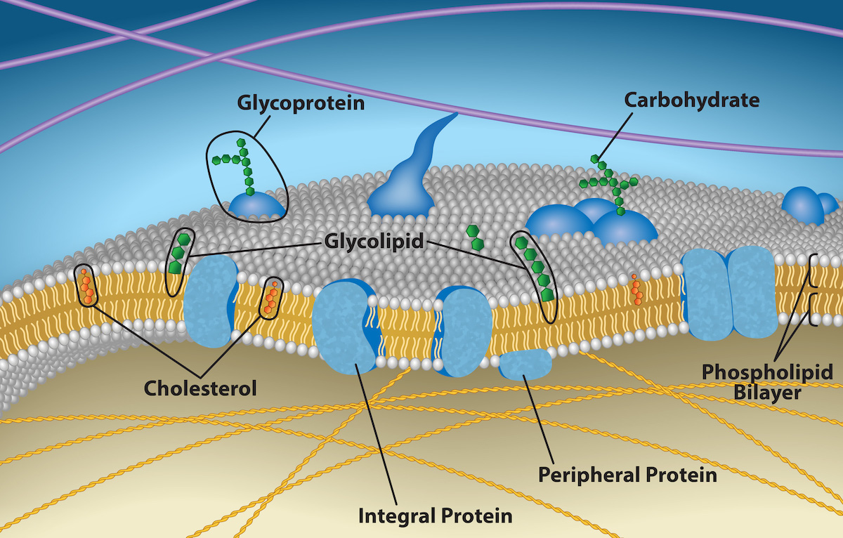
The explanation, the fluid mosaic model, has evolved somewhat over time, but it still best accounts for plasma membrane structure and function as we now understand them. The fluid mosaic model describes the plasma membrane structure as a mosaic of components—including phospholipids, cholesterol, proteins, and carbohydrates—that gives the membrane a fluid character. Plasma membranes range from 5 to 10 nm in thickness. For comparison, human red blood cells, visible via light microscopy, are approximately 8 µm wide, or approximately 1,000 times wider than a plasma membrane. The membrane does look a bit like a sandwich (Figure 5.2).
英語のヒント
この説明である流動モザイクモデルは、時代とともに多少変化してきたが、現在の理解に照らしても、細胞膜の構造と機能を最もよく説明する。流動モザイクモデルでは、細胞膜はリン脂質、コレステロール、タンパク質、糖質などの構成要素が組み合わさったモザイクであり、その構成が膜に流動的な性質を与えるとする。細胞膜の厚さは5〜10 nmである。比較すると、光学顕微鏡で見えるヒトの赤血球の幅は約8 µmで、細胞膜の厚さのおよそ1,000倍である。膜は確かに、少しサンドイッチのように見える(図5.2)。
OpenStax, Rice University; Biology 2e. Credit: Rao, A., Ryan, K., Fletcher, S., Hawkins, A. and Tag, A. Department of Biology, Texas A&M University. · CC BY 4.0 · Source出典

Figure 5.2図 5.2
The plasma membrane fluid mosaic model describes the plasma membrane as a fluid combination of phospholipids, cholesterol, and proteins. Carbohydrates attached to lipids (glycolipids) and to proteins (glycoproteins) extend from the membrane's outward-facing surface. Credit: Rao, A., Ryan, K., Fletcher, S., Hawkins, A. and Tag, A. Department of Biology, Texas A&M University.
流動モザイクモデルは、細胞膜をリン脂質、コレステロール、タンパク質の流動的な組み合わせとして説明する。脂質についた糖質(糖脂質)と、タンパク質についた糖質(糖タンパク質)が、膜の外向きの表面から伸びる。クレジット:A. Rao、K. Ryan、S. Fletcher、A. Hawkins、A. Tag、テキサスA&M大学生物学科。
A plasma membrane's principal components are lipids (phospholipids and cholesterol), proteins, and carbohydrates attached to some of the lipids and proteins. A phospholipid is a molecule consisting of glycerol, two fatty acids, and a phosphate-linked head group. Cholesterol, another lipid comprised of four fused carbon rings, is situated alongside the phospholipids in the membrane's core. The protein, lipid, and carbohydrate proportions in the plasma membrane vary with cell type, but for a typical human cell, protein accounts for about 50 percent of the composition by mass, lipids (of all types) account for about 40 percent, and carbohydrates comprise the remaining 10 percent. However, protein and lipid concentration varies with different cell membranes. For example, myelin, an outgrowth of specialized cells' membrane that insulates the peripheral nerves' axons, contains only 18 percent protein and 76 percent lipid. The mitochondrial inner membrane contains 76 percent protein and only 24 percent lipid. The plasma membrane of human red blood cells is 30 percent lipid. Carbohydrates are present only on the plasma membrane's exterior surface and are attached to proteins, forming glycoproteins, or attached to lipids, forming glycolipids.
英語のヒント
細胞膜の主な構成要素は、脂質(リン脂質とコレステロール)、タンパク質、そして一部の脂質やタンパク質についた糖質である。リン脂質は、グリセロール、2本の脂肪酸、リン酸につながった頭部からなる分子である。別の脂質であるコレステロールは、融合した四つの炭素環からなり、膜の中心部分でリン脂質に沿って位置する。細胞膜内のタンパク質、脂質、糖質の割合は細胞の種類で異なる。典型的なヒトの細胞では、質量比でタンパク質が約50パーセント、全種類を合わせた脂質が約40パーセント、糖質が残りの10パーセントを占める。ただし、タンパク質と脂質の濃度は膜によって異なる。例えば、特殊な細胞の膜が伸びて末梢神経の軸索を絶縁するミエリンは、タンパク質がわずか18パーセント、脂質が76パーセントである。ミトコンドリア内膜ではタンパク質が76パーセント、脂質は24パーセントにすぎない。ヒトの赤血球の細胞膜は30パーセントが脂質である。糖質は細胞膜の外側表面だけに存在し、タンパク質に結合して糖タンパク質を作るか、脂質に結合して糖脂質を作る。