第10章細胞の増殖
10.2 The Cell Cycle10.2 細胞周期
Learning Objectives学習目標
By the end of this section, you will be able to do the following:
この節を学び終えると、次のことができるようになる。
- Describe the three stages of interphase間期の三つの段階を説明する。
- Discuss the behavior of chromosomes during karyokinesis/mitosis核分裂・有糸分裂中の染色体の挙動を論じる。
- Explain how the cytoplasmic content is divided during cytokinesis細胞質分裂で細胞質の内容物が分かれるしくみを説明する。
- Define the quiescent G0 phase静止状態であるG0期を定義する。
The cell cycle is an ordered series of events involving cell growth and cell division that produces two new daughter cells. Cells on the path to cell division proceed through a series of precisely timed and carefully regulated stages of growth, DNA replication, and nuclear and cytoplasmic division that ultimately produces two identical (clone) cells. The cell cycle has two major phases: interphase and the mitotic phase (Figure 10.5). During interphase, the cell grows and DNA is replicated. During the mitotic phase, the replicated DNA and cytoplasmic contents are separated, and the cell cytoplasm is typically partitioned by a third process of the cell cycle called cytokinesis. We should note, however, that interphase and mitosis (karyokinesis) may take place without cytokinesis, in which case cells with multiple nuclei (multinucleate cells) are produced.
英語のヒント
細胞周期は、細胞の成長と分裂にかかわり、新たな2個の娘細胞を作る、順序立った一連の出来事である。分裂へ向かう細胞は、正確に時期を合わせて慎重に調節された成長、DNA複製、核と細胞質の分裂の段階を通り、最終的に同一な2個の細胞(クローン)を生じる。細胞周期には、間期と分裂期という二つの主な時期がある(図10.5)。間期には細胞が成長し、DNAが複製される。分裂期には複製されたDNAと細胞質の内容物が分けられ、通常、細胞周期の第3の過程である細胞質分裂によって細胞質が分割される。ただし、細胞質分裂なしに間期と有糸分裂(核分裂)が進むこともあり、その場合は複数の核を持つ多核細胞が生じる。
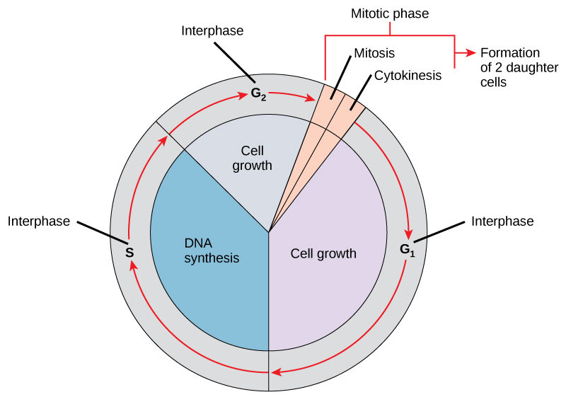
Figure 10.5図 10.5
The cell cycle in multicellular organisms consists of interphase and the mitotic phase. During interphase, the cell grows and the nuclear DNA is duplicated. Interphase is followed by the mitotic phase. During the mitotic phase, the duplicated chromosomes are segregated and distributed into daughter nuclei. Following mitosis, the cytoplasm is usually divided as well by cytokinesis, resulting in two genetically identical daughter cells.
多細胞生物の細胞周期は間期と分裂期からなる。間期には細胞が成長し、核DNAが複製される。その後に分裂期が続く。分裂期には複製された染色体が分離され、娘核へ分配される。有糸分裂に続いて通常は細胞質も細胞質分裂によって分かれ、遺伝的に同一な2個の娘細胞ができる。
Interphase間期
During interphase, the cell undergoes normal growth processes while also preparing for cell division. In order for a cell to move from interphase into the mitotic phase, many internal and external conditions must be met. The three stages of interphase are called G1, S, and G2.
英語のヒント
間期には通常の成長過程が進むとともに、細胞分裂の準備も行われる。間期から分裂期へ進むには、多くの内部条件と外部条件が満たされなければならない。間期の三つの段階をG1期、S期、G2期という。
G1 Phase (First Gap)G1期(第1のギャップ期)
The first stage of interphase is called the G1 phase (first gap) because, from a microscopic point of view, little change is visible. However, during the G1 stage, the cell is quite active at the biochemical level. The cell is accumulating the building blocks of chromosomal DNA and the associated proteins as well as accumulating sufficient energy reserves to complete the task of replicating each chromosome in the nucleus.
英語のヒント
間期の最初の段階はG1期(第1のギャップ期)という。顕微鏡で見る限り、変化がほとんど見えないためである。しかしG1期の細胞は、生化学的には非常に活発である。核内の各染色体の複製を完了するため、染色体DNAや結合タンパク質の材料を蓄えるとともに、十分なエネルギーの備蓄も蓄積する。
S Phase (Synthesis of DNA)S期(DNA合成期)
Throughout interphase, nuclear DNA remains in a semi-condensed chromatin configuration. In the S phase, DNA replication can proceed through the mechanisms that result in the formation of identical pairs of DNA molecules—sister chromatids—that are firmly attached at the centromeric region. The centrosome is also duplicated during the S phase. The two centrosomes will give rise to the mitotic spindle, the apparatus that orchestrates the movement of chromosomes during mitosis. For example, roughly at the center of each animal cell, the centrosomes are associated with a pair of rod-like objects, the centrioles, which are positioned at right angles to each other. Centrioles help organize cell division. We should note, however, that centrioles are not present in the centrosomes of other eukaryotic organisms, such as plants and most fungi.
英語のヒント
間期を通じて、核DNAは半ば凝縮したクロマチンの状態を保つ。S期にはDNA複製が進み、セントロメア領域で強く結び付いた、同一な一対のDNA分子、すなわち姉妹染色分体が形成される。S期には中心体も複製される。二つの中心体は、有糸分裂中の染色体の動きを統率する装置である紡錘体を生み出す。例えば動物細胞のおよそ中央では、中心体は互いに直角に位置する一対の棒状の構造、中心小体と結び付いている。中心小体は細胞分裂の組織化を助ける。ただし植物や大半の菌類など、他の真核生物の中心体には中心小体が存在しないことに注意しよう。
G2 Phase (Second Gap)G2期(第2のギャップ期)
In the G2 phase, the cell replenishes its energy stores and synthesizes proteins necessary for chromosome manipulation and movement. Some cell organelles are duplicated, and the cytoskeleton is dismantled to provide resources for the mitotic phase. There may be additional cell growth during G2. The final preparations for the mitotic phase must be completed before the cell is able to enter the first stage of mitosis.
英語のヒント
G2期にはエネルギーの備蓄を補充し、染色体の操作と移動に必要なタンパク質を合成する。一部の細胞小器官は複製され、細胞骨格は分裂期の資源となるように解体される。G2期にも細胞がさらに成長することがある。有糸分裂の最初の段階へ入る前に、分裂期の最終準備を完了しなければならない。
The Mitotic Phase分裂期
The mitotic phase is a multistep process during which the duplicated chromosomes are aligned, separated, and move into two new, identical daughter cells. The first portion of the mitotic phase is called karyokinesis, or nuclear division. As we have just seen, the second portion of the mitotic phase (and often viewed as a process separate from and following mitosis) is called cytokinesis—the physical separation of the cytoplasmic components into the two daughter cells.
英語のヒント
分裂期は複数の段階からなり、複製された染色体が整列し、分離し、同一な新しい2個の娘細胞へ移動する。分裂期の前半を核分裂という。先に見たように、後半は細胞質分裂と呼ばれ、細胞質の構成要素を物理的に2個の娘細胞へ分ける。この過程は、有糸分裂とは別で、その後に続くものと見なされることも多い。
Karyokinesis (Mitosis)核分裂(有糸分裂)
Karyokinesis, also known as mitosis, is divided into a series of phases—prophase, prometaphase, metaphase, anaphase, and telophase—that result in the division of the cell nucleus (Figure 10.6).
英語のヒント
有糸分裂とも呼ばれる核分裂は、前期、前中期、中期、後期、終期という一連の段階に分かれ、その結果、細胞核が分裂する(図10.6)。
Prophase (the “first phase”): the nuclear envelope starts to dissociate into small vesicles, and the membranous organelles (such as the Golgi complex [Golgi apparatus] and the endoplasmic reticulum), fragment and disperse toward the periphery of the cell. The nucleolus disappears (disperses) as well, and the centrosomes begin to move to opposite poles of the cell. Microtubules that will form the mitotic spindle extend between the centrosomes, pushing them farther apart as the microtubule fibers lengthen. The sister chromatids begin to coil more tightly with the aid of condensin proteins and now become visible under a light microscope.
英語のヒント
前期(「最初の時期」):核膜が小胞へ分かれ始め、ゴルジ複合体(ゴルジ体)や小胞体などの膜性細胞小器官が断片化して、細胞の周辺へ散らばる。核小体も消失して分散し、中心体は細胞の両極へ動き始める。紡錘体を作る微小管が中心体の間を伸び、伸長するにつれて中心体どうしを押し離す。姉妹染色分体はコンデンシンの助けでさらに密に巻き込み、光学顕微鏡で見えるようになる。
Prometaphase (the “first change phase”): Many processes that began in prophase continue to advance. The remnants of the nuclear envelope fragment further, and the mitotic spindle continues to develop as more microtubules assemble and stretch across the length of the former nuclear area. Chromosomes become even more condensed and discrete. Each sister chromatid develops a protein structure called a kinetochore in its centromeric region (Figure 10.7). The proteins of the kinetochore attract and bind to the mitotic spindle microtubules. As the spindle microtubules extend from the centrosomes, some of these microtubules come into contact with and firmly bind to the kinetochores. Once a mitotic fiber attaches to a chromosome, the chromosome will be oriented until the kinetochores of sister chromatids face the opposite poles. Eventually, all the sister chromatids will be attached via their kinetochores to microtubules from opposing poles. Spindle microtubules that do not engage the chromosomes are called polar microtubules. These microtubules overlap each other midway between the two poles and contribute to cell elongation. Astral microtubules are located near the poles, aid in spindle orientation, and are required for the regulation of mitosis.
英語のヒント
前中期(「最初の変化の時期」):前期に始まった多くの過程が進み続ける。核膜の残りはさらに断片化し、より多くの微小管が集合して元の核領域全体に伸びるにつれ、紡錘体の形成が進む。染色体はさらに凝縮して、一つ一つが明確になる。各姉妹染色分体のセントロメア領域には、動原体というタンパク質構造ができる(図10.7)。動原体のタンパク質は紡錘体微小管を引き寄せ、結合する。中心体から紡錘体微小管が伸びると、その一部は動原体に接触して強く結合する。紡錘糸が染色体に付着すると、姉妹染色分体の動原体が反対側の極を向くまで、染色体の向きが調整される。やがてすべての姉妹染色分体が、動原体を介して反対側の極から来る微小管に付着する。染色体に結合しない紡錘体微小管を極間微小管という。これらは両極の中間で重なり合い、細胞の伸長に寄与する。星状微小管は極の近くにあり、紡錘体の向きを整えるのを助け、有糸分裂の調節に必要である。
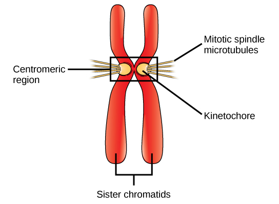


Figure 10.7図 10.7
During prometaphase, mitotic spindle microtubules from opposite poles attach to each sister chromatid at the kinetochore. In anaphase, the connection between the sister chromatids breaks down, and the microtubules pull the chromosomes toward opposite poles.
前中期には、反対側の極から来る紡錘体微小管が、各姉妹染色分体の動原体に付着する。後期には姉妹染色分体の結合が解かれ、微小管が染色体を反対側の極へ引く。
Metaphase (the “change phase”): All the chromosomes are aligned in a plane called the metaphase plate, or the equatorial plane, roughly midway between the two poles of the cell. The sister chromatids are still tightly attached to each other by cohesin proteins. At this time, the chromosomes are maximally condensed.
英語のヒント
中期(「変化の時期」):すべての染色体が、細胞の両極のほぼ中間にある赤道板、または赤道面という平面に並ぶ。姉妹染色分体はまだコヒーシンによって強く結び付いている。この時点で染色体は最大限に凝縮している。
Anaphase (“upward phase”): The cohesin proteins degrade, and the sister chromatids separate at the centromere. Each chromatid, now called a single chromosome, is pulled rapidly toward the centrosome to which its microtubule is attached. The cell becomes visibly elongated (oval shaped) as the polar microtubules slide against each other at the metaphase plate where they overlap.
英語のヒント
後期(「上向きの時期」):コヒーシンが分解され、姉妹染色分体がセントロメアで分離する。各染色分体は今や1本の染色体となり、微小管がつながる中心体へ速やかに引かれる。極間微小管が、互いに重なる赤道板の位置ですれ違うように滑ると、細胞は目に見えて伸び、楕円形になる。
Telophase (the “distance phase”): the chromosomes reach the opposite poles and begin to decondense (unravel), relaxing once again into a stretched-out chromatin configuration. The mitotic spindles are depolymerized into tubulin monomers that will be used to assemble cytoskeletal components for each daughter cell. Nuclear envelopes form around the chromosomes, and nucleosomes appear within the nuclear area.
英語のヒント
終期(「離れた時期」):染色体は反対側の極に達し、凝縮がほどけ始め、再び伸びたクロマチンの状態へ緩む。紡錘体はチューブリン単量体へ脱重合し、それらは各娘細胞の細胞骨格を組み立てるのに使われる。染色体の周囲に核膜が形成され、核領域にヌクレオソームが現れる。