第10章細胞の増殖
Cytokinesis細胞質分裂
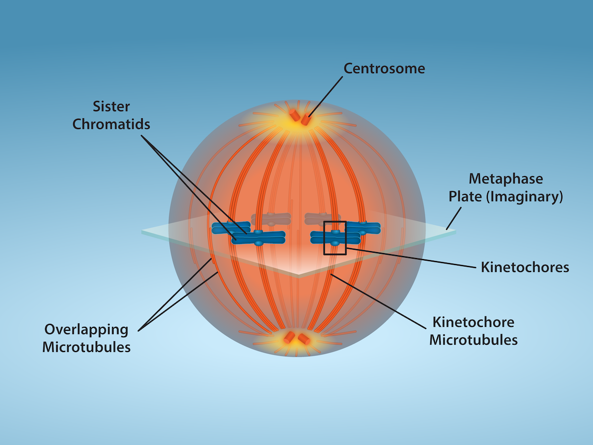
OpenStax, Rice University; Biology 2e. Credit: Fletcher, S., Ryan, K. and Rao, A., Department of Biology, Texas A&M University. · CC BY 4.0 · Source出典

Figure 10.8図 10.8
Mitotic Cell at Metaphase. The microtubule spindle has completed the alignment of chromosomes at the metaphase plate in preparation for the separation of sister chromatids during anaphase. Credit: Fletcher, S., Ryan, K. and Rao, A., Department of Biology, Texas A&M University.
中期の分裂細胞。微小管からなる紡錘体は、後期での姉妹染色分体の分離に備え、赤道板への染色体の整列を完了している。出典:Fletcher, S.、Ryan, K.、Rao, A.、テキサスA&M大学生物学科。
Cytokinesis, or “cell motion,” is sometimes viewed as the second main stage of the mitotic phase, during which cell division is completed via the physical separation of the cytoplasmic components into two daughter cells However, as we have seen earlier, cytokinesis can also be viewed as a separate phase, which may or may not take place following mitosis. If cytokinesis does take place, cell division is not complete until the cell components have been apportioned and completely separated into the two daughter cells. Although the stages of mitosis are similar for most eukaryotes, the process of cytokinesis is quite different for eukaryotes that have cell walls, such as plant cells.
英語のヒント
細胞質分裂は、語源では「細胞の運動」を意味し、分裂期の第2の主要段階と見なされることがある。この段階では細胞質の構成要素を2個の娘細胞へ物理的に分け、細胞分裂を完了する。しかし先に見たとおり、細胞質分裂は有糸分裂の後に起こる場合も起こらない場合もある別の段階と見なすこともできる。細胞質分裂が起こる場合、細胞の成分が2個の娘細胞へ配分され、完全に分離するまで細胞分裂は完了しない。有糸分裂の各段階は大半の真核生物で似ているが、植物細胞のように細胞壁を持つ真核生物では、細胞質分裂の過程がかなり異なる。
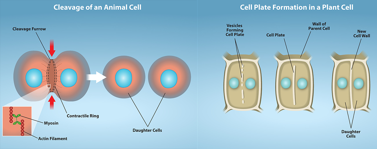
In animal cells, cytokinesis typically starts during late anaphase. A contractile ring composed of actin filaments forms just inside the plasma membrane at the former metaphase plate. The actin filaments pull the equator of the cell inward, forming a fissure. This fissure is called the cleavage furrow. The furrow deepens as the actin ring contracts, and eventually the membrane is cleaved in two (Figure 10.9).
英語のヒント
動物細胞では、細胞質分裂は通常、後期の終わりごろに始まる。アクチンフィラメントからなる収縮環が、元の赤道板の位置で細胞膜のすぐ内側にできる。アクチンフィラメントが細胞の赤道部を内側へ引き、くびれを作る。このくびれを分裂溝という。アクチン環が収縮するにつれて溝が深まり、やがて膜が二つに分かれる(図10.9)。
In plant cells, a new cell wall must form between the daughter cells. During interphase, the Golgi apparatus accumulates enzymes, structural proteins, and glucose molecules prior to breaking into vesicles and dispersing throughout the dividing cell. During telophase, these Golgi vesicles are transported on microtubules to form a phragmoplast (a vesicular structure) at the metaphase plate. There, the vesicles fuse and coalesce from the center toward the cell walls; this structure is called a cell plate. As more vesicles fuse, the cell plate enlarges until it merges with the cell walls at the periphery of the cell. Enzymes use the glucose that has accumulated between the membrane layers to build a new cell wall. The Golgi membranes become parts of the plasma membrane on either side of the new cell wall (Figure 10.9).
英語のヒント
植物細胞では娘細胞の間に新しい細胞壁を作らなければならない。間期のゴルジ体は酵素、構造タンパク質、グルコース分子を蓄え、その後小胞へ分かれて分裂中の細胞全体に散らばる。終期には、そのゴルジ小胞が微小管上を運ばれ、赤道板にフラグモプラスト(小胞状構造)を作る。そこで小胞が融合し、中心から細胞壁へ向けて一体化する。この構造を細胞板という。さらに小胞が融合すると細胞板は広がり、細胞周辺の細胞壁につながる。酵素は膜層の間に蓄積したグルコースで新しい細胞壁を作る。ゴルジ体由来の膜は、新しい細胞壁の両側の細胞膜の一部となる(図10.9)。
OpenStax, Rice University; Biology 2e. Credit: Rao, A., Ryan, K., Hawkins, A.and Fletcher, S. Department of Biology, Texas A&M University. · CC BY 4.0 · Source出典

Figure 10.9図 10.9
Animal cell cleavage - Contraction of actin filaments by myosin pull the “equator” of the cell causing an invagination of the cell (a fissure called a cleavage furrow). The cleavage furrow gets continuously deeper until it eventually divides the cell into two new, independent daughter cells.
Plant cell wall formation - A mixture of enzymes, proteins, and glucose molecules are transported via vesicles to the center of the cell. These vesicles continuously build upon each other until a completely new cell wall has emerged and two new cells are formed, independent of one another.
Credit: Rao, A., Ryan, K., Hawkins, A.and Fletcher, S. Department of Biology, Texas A&M University.
Plant cell wall formation - A mixture of enzymes, proteins, and glucose molecules are transported via vesicles to the center of the cell. These vesicles continuously build upon each other until a completely new cell wall has emerged and two new cells are formed, independent of one another.
Credit: Rao, A., Ryan, K., Hawkins, A.and Fletcher, S. Department of Biology, Texas A&M University.
動物細胞の分割:ミオシンによるアクチンフィラメントの収縮が細胞の「赤道」を引き、細胞を内側へくぼませる。このくびれが分裂溝である。溝は深まり続け、やがて細胞を、互いに独立した2個の新しい娘細胞へ分ける。植物の細胞壁形成:酵素、タンパク質、グルコース分子の混合物が小胞によって細胞中央へ運ばれる。小胞が次々と積み重なり、完全に新しい細胞壁が現れると、互いに独立した2個の細胞ができる。出典:Rao, A.、Ryan, K.、Hawkins, A.、Fletcher, S.、テキサスA&M大学生物学科。
G0 PhaseG0期
Not all cells adhere to the classic cell-cycle pattern in which a newly formed daughter cell immediately enters the preparatory phases of interphase, closely followed by the mitotic phase, and cytokinesis. Cells in G0 phase are not actively preparing to divide. The cell is in a quiescent (inactive) stage that occurs when cells exit the cell cycle. Some cells enter G0 temporarily due to environmental conditions such as availability of nutrients, or stimulation by growth factors. The cell will remain in this phase until conditions improve or until an external signal triggers the onset of G1. Other cells that never or rarely divide, such as mature cardiac muscle and nerve cells, remain in G0 permanently.
英語のヒント
新しくできた娘細胞が直ちに間期の準備段階へ入り、その後すぐに分裂期と細胞質分裂が続くという典型的な細胞周期を、すべての細胞がたどるわけではない。G0期の細胞は積極的に分裂準備をしていない。細胞周期を離れた際に生じる静止(不活性)状態にある。一部の細胞は、栄養素の供給や成長因子による刺激などの環境条件に応じて、一時的にG0期へ入る。条件が改善するか、外部シグナルがG1期の開始を促すまでその状態にとどまる。成熟した心筋細胞や神経細胞のように、まったく、またはほとんど分裂しない細胞は、G0期に永久にとどまる。