第9章細胞間の情報伝達
Cell-Surface Receptors細胞表面受容体
Cell-surface receptors, also known as transmembrane receptors, are cell surface, membrane-anchored (integral) proteins that bind to external ligand molecules. This type of receptor spans the plasma membrane and performs signal transduction, through which an extracellular signal is converted into an intracellular signal. Ligands that interact with cell-surface receptors do not have to enter the cell that they affect. Cell-surface receptors are also called cell-specific proteins or markers because they are specific to individual cell types.
英語のヒント
細胞表面受容体は膜貫通受容体ともいい、細胞の表面で膜に固定された内在性タンパク質で、外部のリガンド分子に結合する。この受容体は細胞膜を貫通しており、細胞外のシグナルを細胞内のシグナルへ変えるシグナル変換を行う。細胞表面受容体と相互作用するリガンドは、作用する細胞の中に入る必要がない。細胞表面受容体は、個々の細胞型に特異的であるため、細胞特異的タンパク質またはマーカーとも呼ばれる。
Because cell-surface receptor proteins are fundamental to normal cell functioning, it should come as no surprise that a malfunction in any one of these proteins could have severe consequences. Errors in the protein structures of certain receptor molecules have been shown to play a role in hypertension (high blood pressure), asthma, heart disease, and cancer.
英語のヒント
細胞表面の受容体タンパク質は細胞が正常に機能するための基礎なので、いずれかの受容体が正常に働かなくなると深刻な結果を招きうることは驚くに当たらない。ある種の受容体分子のタンパク質構造の異常は、高血圧、喘息、心臓病、がんに関与することが示されている。
Each cell-surface receptor has three main components: an external ligand-binding domain called the extracellular domain, a hydrophobic membrane-spanning region called a transmembrane domain, and an intracellular domain inside the cell. The size and extent of each of these domains vary widely, depending on the type of receptor.
英語のヒント
各細胞表面受容体には三つの主な部分がある。細胞外ドメインという外側のリガンド結合領域、膜貫通ドメインという疎水性の膜を横切る領域、そして細胞内にある細胞内ドメインである。それぞれのドメインの大きさや範囲は、受容体の種類によって大きく異なる。
Cell-surface receptors are involved in most of the signaling in multicellular organisms. There are three general categories of cell-surface receptors: ion channel-linked receptors, G-protein-linked receptors, and enzyme-linked receptors.
細胞表面受容体は多細胞生物のシグナル伝達の大部分にかかわる。大きく分けて、イオンチャネル共役型受容体、Gタンパク質共役型受容体、酵素共役型受容体の3種類がある。
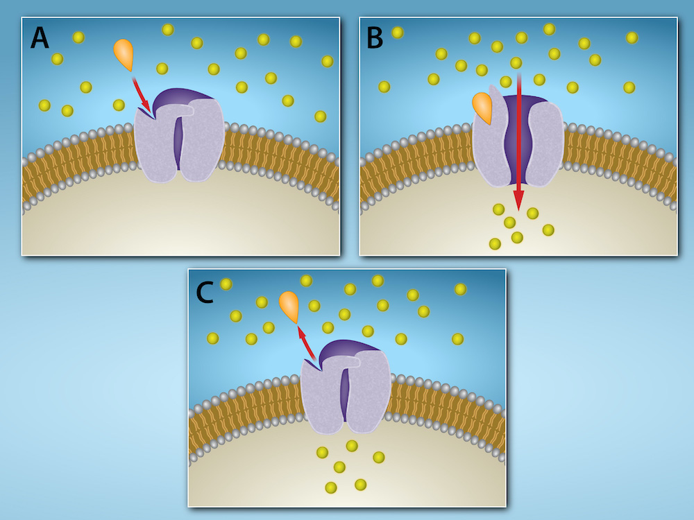
Ion channel-linked receptors bind a ligand and open a channel through the membrane that allows specific ions to pass through. To form a channel, this type of cell-surface receptor has an extensive membrane-spanning region. In order to interact with the double layer of phospholipid fatty acid tails that form the center of the plasma membrane, many of the amino acids in the membrane-spanning region are hydrophobic in nature. Conversely, the amino acids that line the inside of the channel are hydrophilic to allow for the passage of water or ions. When a ligand binds to the extracellular region of the channel, there is a conformational change in the protein's structure that allows ions such as sodium, calcium, magnesium, and hydrogen to pass through (Figure 9.5).
英語のヒント
イオンチャネル共役型受容体はリガンドに結合すると、膜を貫くチャネルを開き、特定のイオンを通す。チャネルを作るため、この種の細胞表面受容体は広い膜貫通領域を持つ。細胞膜の中心をなすリン脂質の脂肪酸尾部の二重層と相互作用するため、膜貫通領域の多くのアミノ酸は疎水性である。逆にチャネルの内側を覆うアミノ酸は、水やイオンを通すために親水性である。リガンドがチャネルの細胞外領域に結合すると、タンパク質の立体構造が変化し、ナトリウム、カルシウム、マグネシウム、水素などのイオンが通れるようになる(図9.5)。
OpenStax, Rice University; Biology 2e. Credit: Rao, A. and Fletcher, S. Department of Biology, Texas A&M University. · CC BY 4.0 · Source出典
Figure 9.5図 9.5
Gated ion channels located in the plasma membrane allow for the controlled flow of ions into and out of the cell. The channel proteins remain closed until a signaling molecule (orange teardrop) binds to the channel protein. Then the channel protein changes conformation and allows ions (yellow circles) to flow into (or out of) the cell. When the signaling molecule is released, the channel protein resumes its closed conformation, preventing ion flow. Credit: Rao, A. and Fletcher, S. Department of Biology, Texas A&M University.
細胞膜にあるゲート付きイオンチャネルは、細胞へのイオンの出入りを制御する。チャネルタンパク質は、シグナル分子(橙色のしずく形)が結合するまでは閉じている。結合すると立体構造が変わり、イオン(黄色の丸)が細胞内へ、または細胞外へ流れる。シグナル分子が離れると閉じた立体構造に戻り、イオンの流れを止める。出典:Rao, A.、Fletcher, S.、テキサスA&M大学生物学科。
G-protein-linked receptors bind a ligand and activate a membrane protein called a G-protein. The activated G-protein then interacts with either an ion channel or an enzyme in the membrane (Figure 9.6). All G-protein-linked receptors have seven transmembrane domains, but each receptor has its own specific extracellular domain and G-protein-binding site.
英語のヒント
Gタンパク質共役型受容体はリガンドに結合し、Gタンパク質という膜タンパク質を活性化する。活性化されたGタンパク質は、膜内のイオンチャネルまたは酵素と相互作用する(図9.6)。すべてのGタンパク質共役型受容体は七つの膜貫通ドメインを持つが、細胞外ドメインとGタンパク質結合部位は受容体ごとに固有である。
Cell signaling using G-protein-linked receptors occurs as a cyclic series of events. Before the ligand binds, the inactive G-protein can bind to a site on the receptor specific for its binding. Once the ligand binds to the receptor, the resulting change in shape activates the G-protein, which releases guanosine diphosphate (GDP) and picks up guanosine 3-phosphate (GTP). The subunits of the G-protein then split into the α subunit and the βγ subunit. One or both of these G-protein fragments may be able to activate other proteins as a result. After awhile, the GTP on the active α subunit of the G-protein is hydrolyzed to GDP and the βγ subunit is deactivated. The subunits reassociate to form the inactive G-protein and the cycle begins anew.
英語のヒント
Gタンパク質共役型受容体による細胞のシグナル伝達は、一連の出来事が循環する形で進む。リガンドが結合する前、不活性なGタンパク質は受容体上の専用の結合部位に結合できる。リガンドが受容体に結合すると、形の変化がGタンパク質を活性化する。Gタンパク質はグアノシン二リン酸(GDP)を放出し、グアノシン三リン酸(GTP)を取り込む。続いて、Gタンパク質のサブユニットがαサブユニットとβγサブユニットに分かれる。その結果、一方または両方の断片が別のタンパク質を活性化できるようになる。しばらくすると、活性型のαサブユニット上のGTPがGDPへ加水分解され、βγサブユニットが不活性化される。サブユニットは再び会合して不活性なGタンパク質となり、サイクルが新たに始まる。
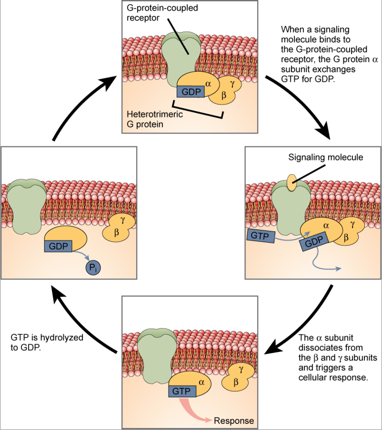


Figure 9.6図 9.6
Heterotrimeric G-proteins have three subunits: α, β, and γ. When a signaling molecule binds to a G-protein-coupled receptor in the plasma membrane, a GDP molecule associated with the α subunit is exchanged for GTP. The β and γ subunits dissociate from the α subunit, and a cellular response is triggered either by the α subunit or the dissociated βγ pair. Hydrolysis of GTP to GDP terminates the signal.
ヘテロ三量体Gタンパク質はα、β、γの三つのサブユニットを持つ。シグナル分子が細胞膜のGタンパク質共役型受容体に結合すると、αサブユニットに結合しているGDPがGTPと交換される。βとγのサブユニットはαから解離し、αサブユニットまたは解離したβγの組が細胞応答を引き起こす。GTPがGDPに加水分解されるとシグナルが終わる。
G-protein-linked receptors have been extensively studied and much has been learned about their roles in maintaining health. Bacteria that are pathogenic to humans can release poisons that interrupt specific G-protein-linked receptor function, leading to illnesses such as pertussis, botulism, and cholera. In cholera (Figure 9.7), for example, the water-borne bacterium Vibrio cholerae produces a toxin, choleragen, that binds to cells lining the small intestine. The toxin then enters these intestinal cells, where it modifies a G-protein that controls the opening of a chloride channel and causes it to remain continuously active, resulting in large losses of fluids from the body and potentially fatal dehydration as a result.
英語のヒント
Gタンパク質共役型受容体は広く研究され、健康を維持する役割について多くのことが分かっている。ヒトに病原性を持つ細菌は、特定のGタンパク質共役型受容体の働きを妨げる毒素を放出することがあり、百日咳、ボツリヌス症、コレラなどを引き起こす。例えばコレラでは(図9.7)、水を介して伝わるコレラ菌Vibrio choleraeが、小腸の内側を覆う細胞に結合するコレラ毒素を作る。毒素はその腸細胞に入り、塩化物イオンチャネルの開口を制御するGタンパク質を変化させて、活性化したままにする。その結果、体から大量の体液が失われ、死に至ることもある脱水が起こる。

OpenStax, Rice University; Biology 2e. (credit: New York City Sanitary Commission) · CC BY 4.0 · Source出典

Figure 9.7図 9.7
Transmitted primarily through contaminated drinking water, cholera is a major cause of death in the developing world and in areas where natural disasters interrupt the availability of clean water. The cholera bacterium, Vibrio cholerae, creates a toxin that modifies G-protein-mediated cell signaling pathways in the intestines. Modern sanitation eliminates the threat of cholera outbreaks, such as the one that swept through New York City in 1866. This poster from that era shows how, at that time, the way that the disease was transmitted was not understood. (credit: New York City Sanitary Commission)
主に汚染された飲料水で伝わるコレラは、発展途上地域や、自然災害で清潔な水が得られなくなった地域における主要な死因である。コレラ菌Vibrio choleraeは、腸内のGタンパク質を介した細胞シグナル伝達経路を変える毒素を作る。近代的な衛生設備は、1866年にニューヨーク市を襲ったようなコレラ流行の脅威をなくす。この時代のポスターからは、当時この病気の伝わり方が理解されていなかったことが分かる。(出典:ニューヨーク市衛生委員会)
Enzyme-linked receptors are cell-surface receptors with intracellular domains that are associated with an enzyme. In some cases, the intracellular domain of the receptor itself is an enzyme. Other enzyme-linked receptors have a small intracellular domain that interacts directly with an enzyme. The enzyme-linked receptors normally have large extracellular and intracellular domains, but the membrane-spanning region consists of a single alpha-helical region of the peptide strand. When a ligand binds to the extracellular domain, a signal is transferred through the membrane, activating the enzyme. Activation of the enzyme sets off a chain of events within the cell that eventually leads to a response. One example of this type of enzyme-linked receptor is the tyrosine kinase receptor (Figure 9.8). A kinase is an enzyme that transfers phosphate groups from ATP to another protein. The tyrosine kinase receptor transfers phosphate groups to tyrosine molecules (tyrosine residues). First, signaling molecules bind to the extracellular domain of two nearby tyrosine kinase receptors. The two neighboring receptors then bond together, or dimerize. Phosphates are then added to tyrosine residues on the intracellular domain of the receptors (phosphorylation). The phosphorylated residues can then transmit the signal to the next messenger within the cytoplasm.
英語のヒント
酵素共役型受容体は、酵素と結び付いた細胞内ドメインを持つ細胞表面受容体である。細胞内ドメイン自体が酵素である場合もあれば、小さな細胞内ドメインが酵素と直接相互作用する場合もある。通常、細胞外ドメインと細胞内ドメインは大きいが、膜貫通領域はペプチド鎖の一つのαらせん領域からなる。リガンドが細胞外ドメインに結合すると、膜を介してシグナルが伝わり、酵素が活性化される。活性化した酵素は細胞内で一連の出来事を起こし、最終的に応答へつながる。この種の受容体の一例がチロシンキナーゼ受容体である(図9.8)。キナーゼはATPから別のタンパク質へリン酸基を移す酵素である。チロシンキナーゼ受容体はチロシン分子(チロシン残基)へリン酸基を移す。まずシグナル分子が、近くにある二つのチロシンキナーゼ受容体の細胞外ドメインに結合する。次に隣接する受容体どうしが結合し、二量体化する。その後、受容体の細胞内ドメインのチロシン残基へリン酸基が付加される(リン酸化)。リン酸化された残基は、細胞質内の次の伝達物質へシグナルを伝えられる。