第41章浸透圧調節と排泄
41.3 Excretion Systems41.3 排泄系
Learning Objectives学習目標
By the end of this section, you will be able to do the following:
この節を学ぶと、次のことができるようになる。
- Explain how vacuoles, present in microorganisms, work to excrete waste微生物の液胞が老廃物を排泄する仕組みを説明する。
- Describe the way in which flame cells and nephridia in worms perform excretory functions and maintain osmotic balance蠕虫の炎細胞と腎管が排泄機能を果たし、浸透圧平衡を保つ仕組みを説明する。
- Explain how insects use Malpighian tubules to excrete wastes and maintain osmotic balance昆虫がマルピーギ管を使って老廃物を排泄し、浸透圧平衡を保つ仕組みを説明する。
Microorganisms and invertebrate animals use more primitive and simple mechanisms to get rid of their metabolic wastes than the mammalian system of kidney and urinary function. Three excretory systems evolved in organisms before complex kidneys: vacuoles, flame cells, and Malpighian tubules.
英語のヒント
微生物と無脊椎動物は、哺乳類の腎臓と排尿機能よりも原始的で単純な仕組みを使って、代謝老廃物を除いている。複雑な腎臓が現れる前に、生物では液胞、炎細胞、マルピーギ管という3つの排泄系が進化した。
Contractile Vacuoles in Microorganisms微生物の収縮胞
The most fundamental feature of life is the presence of a cell. In other words, a cell is the simplest functional unit of a life. Bacteria are unicellular, prokaryotic organisms that have some of the least complex life processes in place; however, prokaryotes such as bacteria do not contain membrane-bound vacuoles. The cells of microorganisms like bacteria, protozoa, and fungi are bound by cell membranes and use them to interact with the environment. Some cells, including some leucocytes in humans, are able to engulf food by endocytosis—the formation of vesicles by involution of the cell membrane within the cells. The same vesicles are able to interact and exchange metabolites with the intracellular environment. In some unicellular eukaryotic organisms such as the amoeba, shown in Figure 41.9, cellular wastes and excess water are excreted by exocytosis, when the contractile vacuoles merge with the cell membrane and expel wastes into the environment. Contractile vacuoles (CV) should not be confused with vacuoles, which store food or water.
英語のヒント
生命の最も基本的な特徴は、細胞が存在することである。言い換えると、細胞は生命の最も単純な機能単位である。細菌は単細胞の原核生物で、最も単純な部類の生命活動を営む。しかし、細菌などの原核生物は、膜で囲まれた液胞をもたない。細菌、原生動物、菌類などの微生物の細胞は細胞膜に囲まれ、その膜を使って環境とやり取りする。ヒトの一部の白血球などには、エンドサイトーシスによって食物を取り込める細胞がある。これは、細胞膜が細胞の内側へ陥入して小胞を形成する過程である。同じ小胞は、細胞内の環境と相互作用し、代謝産物を交換できる。図41.9のアメーバなど、一部の単細胞真核生物では、細胞の老廃物と余分な水はエキソサイトーシスによって排泄される。収縮胞が細胞膜と融合し、老廃物を外の環境へ排出するのである。収縮胞(CV)を、食物や水を蓄える液胞と混同してはいけない。
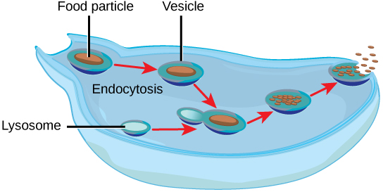
Figure 41.9図 41.9
Some unicellular organisms, such as the amoeba, ingest food by endocytosis. The food vesicle fuses with a lysosome, which digests the food. Waste is excreted by exocytosis.
アメーバなどの単細胞生物は、エンドサイトーシスで食物を取り込む。食物を含む小胞はリソソームと融合し、食物が消化される。老廃物はエキソサイトーシスで排泄される。
Flame Cells of Planaria and Nephridia of Wormsプラナリアの炎細胞と蠕虫の腎管
As multicellular systems evolved to have organ systems that divided the metabolic needs of the body, individual organs evolved to perform the excretory function. Planaria are flatworms that live in freshwater. Their excretory system consists of two tubules connected to a highly branched duct system. The cells in the tubules are called flame cells (or protonephridia) because they have a cluster of cilia that looks like a flickering flame when viewed under the microscope, as illustrated in Figure 41.10a. The cilia propel waste matter down the tubules and out of the body through excretory pores that open on the body surface; cilia also draw water from the interstitial fluid, allowing for filtration. Any valuable metabolites are recovered by reabsorption. Flame cells are found in flatworms, including parasitic tapeworms and free-living planaria. They also maintain the organism’s osmotic balance.
英語のヒント
多細胞生物が進化し、体の代謝上の必要を分担する器官系をもつようになると、排泄機能を果たす個々の器官も進化した。プラナリアは淡水に生息する扁形動物である。その排泄系は、高度に枝分かれした管系につながる2本の細管からなる。細管内の細胞は炎細胞(または原腎管)と呼ばれる。図41.10aのように、顕微鏡で見ると揺らめく炎のように見える繊毛の束をもつためである。繊毛は老廃物を細管に沿って押し流し、体表に開く排泄孔から体外へ出す。また、繊毛は間質液から水を引き込み、濾過を可能にする。有用な代謝産物は再吸収によって回収される。炎細胞は、寄生性の条虫や自由生活性のプラナリアなど、扁形動物に見られる。浸透圧平衡を保つ役割も果たす。
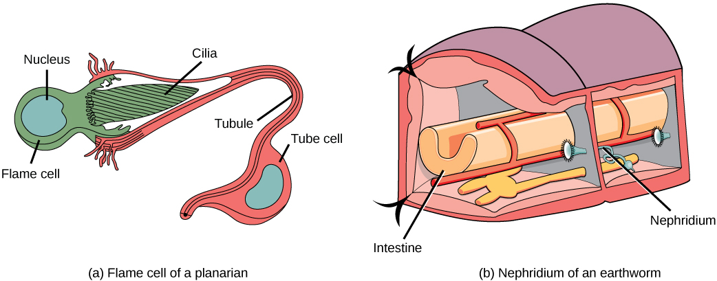
Figure 41.10図 41.10
In the excretory system of the (a) planaria, cilia of flame cells propel waste through a tubule formed by a tube cell. Tubules are connected into branched structures that lead to pores located all along the sides of the body. The filtrate is secreted through these pores. In (b) annelids such as earthworms, nephridia filter fluid from the coelom, or body cavity. Beating cilia at the opening of the nephridium draw water from the coelom into a tubule. As the filtrate passes down the tubules, nutrients and other solutes are reabsorbed by capillaries. Filtered fluid containing nitrogenous and other wastes is stored in a bladder and then secreted through a pore in the side of the body.
(a)プラナリアの排泄系では、炎細胞の繊毛が、管細胞の作る細管に老廃物を押し流す。細管は枝分かれした構造につながり、体の側面全体に並ぶ孔へ通じる。濾液はこれらの孔から分泌される。(b)ミミズなどの環形動物では、腎管が体腔の液を濾過する。腎管の開口部で動く繊毛が、体腔から細管へ水を引き込む。濾液が細管を進む間に、栄養素や他の溶質が毛細血管によって再吸収される。窒素性老廃物などを含む濾過された液は膀胱に蓄えられ、その後、体の側面の孔から分泌される。
Earthworms (annelids) have slightly more evolved excretory structures called nephridia, illustrated in Figure 41.10b. A pair of nephridia is present on each segment of the earthworm. They are similar to flame cells in that they have a tubule with cilia. Excretion occurs through a pore called the nephridiopore. They are more evolved than the flame cells in that they have a system for tubular reabsorption by a capillary network before excretion.
英語のヒント
ミミズ(環形動物)は、図41.10bに示す腎管という、やや進化した排泄構造をもつ。各体節に1対の腎管がある。繊毛のある細管をもつ点では、炎細胞と似ている。排泄は腎管孔と呼ばれる孔を通じて行われる。排泄前に毛細血管網による尿細管再吸収を行う仕組みがある点で、炎細胞より進化している。
Malpighian Tubules of Insects昆虫のマルピーギ管
Malpighian tubules are found lining the gut of some species of arthropods, such as the bee illustrated in Figure 41.11. They are usually found in pairs and the number of tubules varies with the species of insect. Malpighian tubules are convoluted, which increases their surface area, and they are lined with microvilli for reabsorption and maintenance of osmotic balance. Malpighian tubules work cooperatively with specialized glands in the wall of the rectum. Body fluids are not filtered as in the case of nephridia; urine is produced by tubular secretion mechanisms by the cells lining the Malpighian tubules that are bathed in hemolymph (a mixture of blood and interstitial fluid that is found in insects and other arthropods as well as most mollusks). Metabolic wastes like uric acid freely diffuse into the tubules. There are exchange pumps lining the tubules, which actively transport H+ ions into the cell and K+ or Na+ ions out; water passively follows to form urine. The secretion of ions alters the osmotic pressure which draws water, electrolytes, and nitrogenous waste (uric acid) into the tubules. Water and electrolytes are reabsorbed when these organisms are faced with low-water environments, and uric acid is excreted as a thick paste or powder. Not dissolving wastes in water helps these organisms to conserve water; this is especially important for life in dry environments.
英語のヒント
マルピーギ管は、図41.11のハチのような一部の節足動物で、消化管に沿って見られる。通常は対になっており、管の数は昆虫の種によって異なる。マルピーギ管は曲がりくねって表面積を増やしており、内面には再吸収と浸透圧平衡の維持のための微絨毛が並ぶ。また、直腸の壁にある特殊な腺と協力して働く。腎管の場合とは異なり、体液は濾過されない。尿は、血リンパに浸されたマルピーギ管内面の細胞による尿細管分泌の仕組みで作られる。血リンパは血液と間質液の混合物で、昆虫や他の節足動物、および大部分の軟体動物に見られる。尿酸などの代謝老廃物は、管内へ自由に拡散する。管の内面には交換ポンプがあり、H+を細胞内へ、K+またはNa+を細胞外へ能動輸送する。水は受動的に後に続き、尿が形成される。イオンの分泌によって浸透圧が変わり、水、電解質、窒素性老廃物(尿酸)が管内へ引き込まれる。これらの生物が水の少ない環境に置かれると、水と電解質は再吸収され、尿酸は濃いペースト状または粉末状で排泄される。老廃物を水に溶かさないことは、水の節約に役立つ。乾燥した環境で生きるうえで、これは特に重要である。
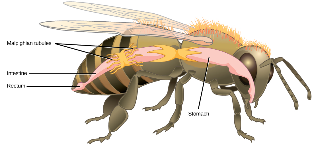


Figure 41.11図 41.11
Malpighian tubules of insects and other terrestrial arthropods remove nitrogenous wastes and other solutes from the hemolymph. Na+ and/or K+ ions are actively transported into the lumen of the tubules. Water then enters the tubules via osmosis, forming urine. The urine passes through the intestine, and into the rectum. There, nutrients diffuse back into the hemolymph. Na+ and/or K+ ions are pumped into the hemolymph, and water follows. The concentrated waste is then excreted.
昆虫などの陸生節足動物のマルピーギ管は、血リンパから窒素性老廃物やその他の溶質を除く。Na+やK+が管腔へ能動輸送され、続いて浸透によって水が管内に入り、尿が作られる。尿は腸を通り、直腸に入る。そこで栄養素が血リンパへ拡散して戻る。Na+やK+も血リンパへポンプ輸送され、水が後に続く。その後、濃縮された老廃物が排泄される。