第41章浸透圧調節と排泄
41.2 The Kidneys and Osmoregulatory Organs41.2 腎臓と浸透圧調節器官
Learning Objectives学習目標
By the end of this section, you will be able to do the following:
この節を学ぶと、次のことができるようになる。
- Explain how the kidneys serve as the main osmoregulatory organs in mammalian systems哺乳類で腎臓が主要な浸透圧調節器官としてどのように働くかを説明する。
- Describe the structure of the kidneys and the functions of the parts of the kidney腎臓の構造と、各部の機能を説明する。
- Describe how the nephron is the functional unit of the kidney and explain how it actively filters blood and generates urineネフロンが腎臓の機能単位であることを説明し、血液を活発に濾過して尿を作る仕組みを述べる。
- Detail the three steps in the formation of urine: glomerular filtration, tubular reabsorption, and tubular secretion尿形成の3段階である糸球体濾過、尿細管再吸収、尿細管分泌を詳しく説明する。
Although the kidneys are the major osmoregulatory organ, the skin and lungs also play a role in the process. Water and electrolytes are lost through sweat glands in the skin, which helps moisturize and cool the skin surface, while the lungs expel a small amount of water in the form of mucous secretions and via evaporation of water vapor.
英語のヒント
腎臓が主要な浸透圧調節器官であるが、皮膚と肺もこの過程に関わっている。皮膚の汗腺からは水と電解質が失われ、それが皮膚の表面を潤し、冷やすのに役立つ。一方、肺は粘液の分泌や水蒸気の蒸発によって、少量の水を排出する。
Kidneys: The Main Osmoregulatory Organ腎臓:主要な浸透圧調節器官
The kidneys, illustrated in Figure 41.4, are a pair of bean-shaped structures that are located just below and posterior to the liver in the peritoneal cavity. The adrenal glands sit on top of each kidney and are also called the suprarenal glands. Kidneys filter blood and purify it. All the blood in the human body is filtered many times a day by the kidneys; these organs use up almost 25 percent of the oxygen absorbed through the lungs to perform this function. Oxygen allows the kidney cells to efficiently manufacture chemical energy in the form of ATP through aerobic respiration. The filtrate coming out of the kidneys is called urine.
英語のヒント
図41.4に示す腎臓は、腹膜腔内で肝臓のすぐ下かつ後方に位置する、一対の豆形の構造である。それぞれの腎臓の上には副腎があり、英語ではadrenal glandsのほかsuprarenal glandsとも呼ばれる。腎臓は血液を濾過して浄化する。人体の血液はすべて、1日に何度も腎臓で濾過される。腎臓はこの機能を果たすために、肺で吸収される酸素のほぼ25%を消費する。酸素によって、腎臓の細胞は好気呼吸を通じてATPという形の化学エネルギーを効率よく作れる。腎臓から出てくる濾液を尿という。
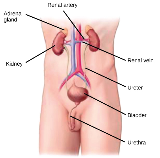

Figure 41.4図 41.4
Kidneys filter the blood, producing urine that is stored in the bladder prior to elimination through the urethra. (credit: modification of work by NCI)
腎臓は血液を濾過して尿を作る。尿は膀胱に蓄えられたのち、尿道を通って排出される。(出典:NCIの作品を改変)
Kidney Structure腎臓の構造
Externally, the kidneys are surrounded by three layers, illustrated in Figure 41.5. The outermost layer is a tough connective tissue layer called the renal fascia. The second layer is called the perirenal fat capsule, which helps anchor the kidneys in place. The third and innermost layer is the renal capsule. Internally, the kidney has three regions—an outer cortex, a medulla in the middle, and the renal pelvis in the region called the hilum of the kidney. The hilum is the concave part of the bean-shape where blood vessels and nerves enter and exit the kidney; it is also the point of exit for the ureters. The renal cortex is granular due to the presence of nephrons—the functional unit of the kidney. The medulla consists of multiple pyramidal tissue masses, called the renal pyramids. In between the pyramids are spaces called renal columns through which the blood vessels pass. The tips of the pyramids, called renal papillae, point toward the renal pelvis. There are, on average, eight renal pyramids in each kidney. The renal pyramids along with the adjoining cortical region are called the lobes of the kidney. The renal pelvis leads to the ureter on the outside of the kidney. On the inside of the kidney, the renal pelvis branches out into two or three extensions called the major calyces, which further branch into the minor calyces. The ureters are urine-bearing tubes that exit the kidney and empty into the urinary bladder.
英語のヒント
腎臓の外側は、図41.5のように3層で囲まれている。最外層は、腎筋膜という丈夫な結合組織の層である。第2層は腎周囲脂肪被膜といい、腎臓を所定の位置に固定するのに役立つ。第3層である最内層は、腎被膜である。腎臓の内部には、外側の皮質、中間の髄質、そして腎門と呼ばれる領域にある腎盂という3つの領域がある。腎門は豆形のくぼんだ部分で、血管や神経が出入りする。また、尿管が出る場所でもある。腎皮質は、腎臓の機能単位であるネフロンが存在するため、粒状に見える。髄質は、腎錐体という複数の錐体状の組織塊からなる。錐体の間には腎柱という部分があり、そこを血管が通る。錐体の先端は腎乳頭と呼ばれ、腎盂に向かっている。腎錐体は、各腎臓に平均8個ある。腎錐体とそれに隣接する皮質の領域を合わせて腎葉という。腎盂は腎臓の外で尿管につながる。腎臓の内側では、腎盂は大腎杯という2〜3個の突出部に枝分かれし、そこからさらに小腎杯へ分かれる。尿管は尿を運ぶ管で、腎臓から出て膀胱へ内容物を流し込む。
Because the kidney filters blood, its network of blood vessels is an important component of its structure and function. The arteries, veins, and nerves that supply the kidney enter and exit at the renal hilum. Renal blood supply starts with the branching of the aorta into the renal arteries (which are each named based on the region of the kidney they pass through) and ends with the exiting of the renal veins to join the inferior vena cava. The renal arteries split into several segmental arteries upon entering the kidneys. Each segmental artery splits further into several interlobar arteries and enters the renal columns, which supply the renal lobes. The interlobar arteries split at the junction of the renal cortex and medulla to form the arcuate arteries. The arcuate “bow shaped” arteries form arcs along the base of the medullary pyramids. Cortical radiate arteries, as the name suggests, radiate out from the arcuate arteries. The cortical radiate arteries branch into numerous afferent arterioles, and then enter the capillaries supplying the nephrons. Veins trace the path of the arteries and have similar names, except there are no segmental veins.
英語のヒント
腎臓は血液を濾過するので、血管網はその構造と機能の重要な構成要素である。腎臓へ通じる動脈、静脈、神経は、腎門で出入りする。腎臓の血流は、大動脈が腎動脈(腎臓内で通過する領域に応じて、それぞれ名付けられる)へ分岐するところから始まり、腎静脈が出て下大静脈に合流するところで終わる。腎動脈は腎臓に入ると、数本の区域動脈に分かれる。各区域動脈はさらに数本の葉間動脈に分かれ、腎柱に入って腎葉に血液を送る。葉間動脈は腎皮質と髄質の境界で分かれ、弓状動脈を作る。弓状、つまり「弓形」の動脈は、髄質の錐体の底部に沿って弧を描く。皮質放線動脈はその名のとおり、弓状動脈から放射状に伸びる。皮質放線動脈は多数の輸入細動脈に枝分かれし、その先でネフロンに血液を送る毛細血管に入る。静脈は動脈の経路をたどり、似た名前が付いているが、区域静脈は存在しない。
As mentioned previously, the functional unit of the kidney is the nephron, illustrated in Figure 41.6. Each kidney is made up of over one million nephrons that dot the renal cortex, giving it a granular appearance when sectioned sagittally. There are two types of nephrons—cortical nephrons (85 percent), which are deep in the renal cortex, and juxtamedullary nephrons (15 percent), which lie in the renal cortex close to the renal medulla. A nephron consists of three parts—a renal corpuscle, a renal tubule, and the associated capillary network, which originates from the cortical radiate arteries.
英語のヒント
先に述べたように、腎臓の機能単位は、図41.6に示すネフロンである。各腎臓は100万個を超えるネフロンからなり、それらが腎皮質に点在するため、矢状断にすると粒状に見える。ネフロンには、腎皮質の深部にある皮質ネフロン(85%)と、腎髄質に近い腎皮質にある傍髄質ネフロン(15%)の2種類がある。ネフロンは、腎小体、尿細管、および皮質放線動脈から生じる付随した毛細血管網という3つの部分で構成される。