第14章DNAの構造と機能
14.5 DNA Replication in Eukaryotes14.5 真核生物のDNA複製
Learning Objectives学習目標
By the end of this section, you will be able to do the following:
この節を学ぶと、次のことができるようになる。
- Discuss the similarities and differences between DNA replication in eukaryotes and prokaryotes真核生物と原核生物のDNA複製の共通点と相違点について論じる。
- State the role of telomerase in DNA replicationDNA複製におけるテロメラーゼの役割を述べる。
Eukaryotic genomes are much more complex and larger in size than prokaryotic genomes. Eukaryotes also have a number of different linear chromosomes. The human genome has 3 billion base pairs per haploid set of chromosomes, and 6 billion base pairs are replicated during the S phase of the cell cycle. There are multiple origins of replication on each eukaryotic chromosome; humans can have up to 100,000 origins of replication across the genome. The rate of replication is approximately 100 nucleotides per second, much slower than prokaryotic replication. In yeast, which is a eukaryote, special sequences known as autonomously replicating sequences (ARS) are found on the chromosomes. These are equivalent to the origin of replication in E. coli.
英語のヒント
真核生物のゲノムは、原核生物のゲノムよりはるかに複雑で大きい。また、真核生物は複数の異なる直鎖状染色体をもつ。ヒトのゲノムには、染色体の半数体1組あたり30億塩基対があり、細胞周期のS期には60億塩基対が複製される。各真核生物染色体には複数の複製起点があり、ヒトではゲノム全体で最大10万か所に達する。複製速度は1秒あたり約100ヌクレオチドで、原核生物の複製よりはるかに遅い。真核生物である酵母の染色体には、自律複製配列(ARS)という特別な配列がある。これは、大腸菌の複製起点に相当する。
The number of DNA polymerases in eukaryotes is much more than in prokaryotes: 14 are known, of which five are known to have major roles during replication and have been well studied. They are known as pol α, pol β, pol γ, pol δ, and pol ε.
英語のヒント
真核生物のDNAポリメラーゼの種類は、原核生物よりはるかに多い。14種類が知られ、そのうち5種類は複製で主要な役割を担うことがわかっており、よく研究されている。それらはpol α、pol β、pol γ、pol δ、pol εと呼ばれる。
The essential steps of replication are the same as in prokaryotes. Before replication can start, the DNA has to be made available as a template. Eukaryotic DNA is bound to basic proteins known as histones to form structures called nucleosomes. Histones must be removed and then replaced during the replication process, which helps to account for the lower replication rate in eukaryotes. The chromatin (the complex between DNA and proteins) may undergo some chemical modifications, so that the DNA may be able to slide off the proteins or be accessible to the enzymes of the DNA replication machinery. At the origin of replication, a pre-replication complex is made with other initiator proteins. Helicase and other proteins are then recruited to start the replication process (Table 14.2).
英語のヒント
複製の基本的な段階は、原核生物の場合と同じである。複製を開始する前に、DNAを鋳型として利用できる状態にしなければならない。真核生物のDNAは、ヒストンという塩基性タンパク質に結合して、ヌクレオソームという構造を形成している。複製の過程では、ヒストンをいったん取り除き、その後で元に戻す必要がある。これが、真核生物の複製速度が遅い理由の1つである。クロマチン(DNAとタンパク質の複合体)には化学修飾が加わることがあり、その結果、DNAがタンパク質から外れたり、DNA複製を担う酵素が近づけたりするようになる。複製起点には、ほかの開始タンパク質とともに複製前複合体が作られる。その後、ヘリカーゼなどのタンパク質が集められ、複製が始まる(表14.2)。
Difference between Prokaryotic and Eukaryotic Replication 原核生物と真核生物の複製の違い |
||
|---|---|---|
Property 性質 |
Prokaryotes 原核生物 |
Eukaryotes 真核生物 |
Origin of replication 複製起点 |
Single 1か所 |
Multiple 複数 |
Rate of replication 複製速度 |
1000 nucleotides/s 1,000ヌクレオチド/秒 |
50 to 100 nucleotides/s 50〜100ヌクレオチド/秒 |
DNA polymerase types DNAポリメラーゼの種類 |
5 |
14 |
Telomerase テロメラーゼ |
Not present 存在しない |
Present 存在する |
RNA primer removal RNAプライマーの除去 |
DNA pol I |
RNase H |
Strand elongation 鎖の伸長 |
DNA pol III |
Pol α, pol δ, pol ε Pol α、pol δ、pol ε |
Sliding clamp スライディングクランプ |
Sliding clamp スライディングクランプ |
PCNA |
A helicase using the energy from ATP hydrolysis opens up the DNA helix. Replication forks are formed at each replication origin as the DNA unwinds. The opening of the double helix causes over-winding, or supercoiling, in the DNA ahead of the replication fork. These are resolved with the action of topoisomerases. Primers are formed by the enzyme primase, and using the primer, DNA pol can start synthesis. Three major DNA polymerases are then involved: α, δ and ε. DNA pol α adds a short (20 to 30 nucleotides) DNA fragment to the RNA primer on both strands, and then hands off to a second polymerase. While the leading strand is continuously synthesized by the enzyme pol ε, the lagging strand is synthesized by pol δ. A sliding clamp protein known as PCNA (proliferating cell nuclear antigen) holds the DNA pol in place so that it does not slide off the DNA. As pol δ runs into the primer RNA on the lagging strand, it displaces it from the DNA template. The displaced primer RNA is then removed by RNase H (AKA flap endonuclease) and replaced with DNA nucleotides. The Okazaki fragments in the lagging strand are joined after the replacement of the RNA primers with DNA. The gaps that remain are sealed by DNA ligase, which forms the phosphodiester bond.
英語のヒント
ヘリカーゼはATPの加水分解によるエネルギーを使ってDNAらせんを開く。DNAがほどけるにつれて、各複製起点に複製フォークが形成される。二重らせんが開くと、複製フォークの前方のDNAには過剰な巻き込み、すなわち超らせんが生じる。これをトポイソメラーゼの作用が解消する。プライマーゼという酵素がプライマーを作り、DNAポリメラーゼはそれを用いて合成を開始できる。続いて、α、δ、εという3つの主要なDNAポリメラーゼが関与する。DNA pol αは、両方の鎖のRNAプライマーに短いDNA断片(20〜30ヌクレオチド)を付加し、その後、別のポリメラーゼに引き継ぐ。リーディング鎖はpol εによって連続的に合成されるが、ラギング鎖はpol δによって合成される。PCNA(増殖細胞核抗原)というスライディングクランプタンパク質がDNAポリメラーゼを保持し、DNAから外れないようにする。pol δがラギング鎖上のRNAプライマーに達すると、プライマーをDNA鋳型から押しのける。押しのけられたRNAプライマーは、RNase H(別名フラップエンドヌクレアーゼ)によって除去され、DNAヌクレオチドに置き換えられる。ラギング鎖の岡崎フラグメントは、RNAプライマーがDNAに置き換わった後でつながれる。残った隙間は、ホスホジエステル結合を形成するDNAリガーゼによって閉じられる。
Telomere replicationテロメアの複製
Unlike prokaryotic chromosomes, eukaryotic chromosomes are linear. As you’ve learned, the enzyme DNA pol can add nucleotides only in the 5' to 3' direction. In the leading strand, synthesis continues until the end of the chromosome is reached. On the lagging strand, DNA is synthesized in short stretches, each of which is initiated by a separate primer. When the replication fork reaches the end of the linear chromosome, there is no way to replace the primer on the 5’ end of the lagging strand. The DNA at the ends of the chromosome thus remains unpaired, and over time these ends, called telomeres, may get progressively shorter as cells continue to divide.
英語のヒント
原核生物の染色体と異なり、真核生物の染色体は直鎖状である。すでに学んだように、DNAポリメラーゼは5′から3′の方向にしかヌクレオチドを付加できない。リーディング鎖では、染色体の末端に達するまで合成が続く。ラギング鎖ではDNAは短い断片として合成され、それぞれが別々のプライマーによって開始される。複製フォークが直鎖状染色体の末端に達すると、ラギング鎖の5′末端にあるプライマーを置き換える方法がない。そのため、染色体末端のDNAは対を作らないまま残る。細胞分裂が続くにつれて、テロメアと呼ばれるこの末端は、時間とともに少しずつ短くなることがある。
Telomeres comprise repetitive sequences that code for no particular gene. In humans, a six-base-pair sequence, TTAGGG, is repeated 100 to 1000 times in the telomere regions. In a way, these telomeres protect the genes from getting deleted as cells continue to divide. The telomeres are added to the ends of chromosomes by a separate enzyme, telomerase (Figure 14.16), whose discovery helped in the understanding of how these repetitive chromosome ends are maintained. The telomerase enzyme contains a catalytic part and a built-in RNA template. It attaches to the end of the chromosome, and DNA nucleotides complementary to the RNA template are added on the 3' end of the DNA strand. Once the 3' end of the lagging strand template is sufficiently elongated, DNA polymerase can add the nucleotides complementary to the ends of the chromosomes. Thus, the ends of the chromosomes are replicated.
英語のヒント
テロメアは、特定の遺伝子をコードしない反復配列からなる。ヒトのテロメア領域では、TTAGGGという6塩基対の配列が100〜1,000回繰り返されている。ある意味では、このテロメアが、細胞分裂を繰り返す際に遺伝子が失われるのを防いでいる。テロメアは、別の酵素であるテロメラーゼによって染色体の末端に付加される(図14.16)。テロメラーゼの発見は、この反復配列をもつ染色体末端がどのように維持されるかを理解するのに役立った。テロメラーゼには、触媒として働く部分と、内蔵されたRNA鋳型がある。テロメラーゼが染色体末端に結合すると、RNA鋳型に相補的なDNAヌクレオチドが、DNA鎖の3′末端に付加される。ラギング鎖の鋳型の3′末端が十分に伸びると、DNAポリメラーゼが染色体末端に相補的なヌクレオチドを付加できるようになる。こうして、染色体の末端が複製される。
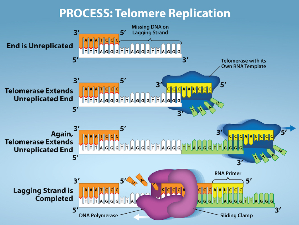
OpenStax, Rice University; Biology 2e. Credit: Rao, A. and Fletcher, S. Department of Biology, Texas A&M University. · CC BY 4.0 · Source出典
Figure 14.15図 14.15
The ends of linear chromosomes are maintained by the action of the telomerase enzyme. Credit: Rao, A. and Fletcher, S. Department of Biology, Texas A&M University.
直鎖状染色体の末端は、テロメラーゼという酵素の作用によって維持される。クレジット:Rao, A. and Fletcher, S.、テキサスA&M大学生物学科。
Telomerase is typically active in germ cells and adult stem cells. It is not active in adult somatic cells. For their discovery of telomerase and its action, Elizabeth Blackburn, Carol W. Greider, and Jack W. Szostak (Figure 14.16) received the Nobel Prize for Medicine and Physiology in 2009. Later research using HeLa cells (obtained from Henrietta Lacks) confirmed that telomerase is present in human cells. And in 2001, researchers including Diane L. Wright found that telomerase is necessary for cells in human embryos to rapidly proliferate.
英語のヒント
テロメラーゼは通常、生殖細胞と成体幹細胞で活性を示す。成体の体細胞では活性を示さない。テロメラーゼとその働きを発見した功績により、エリザベス・ブラックバーン、キャロル・W・グライダー、ジャック・W・ショスタク(図14.16)は、2009年にノーベル医学生理学賞を受賞した。その後、HeLa細胞(ヘンリエッタ・ラックスから採取された細胞)を使った研究により、ヒトの細胞にもテロメラーゼが存在することが確認された。また2001年には、ダイアン・L・ライトらの研究者が、ヒト胚の細胞が急速に増殖するにはテロメラーゼが必要であることを見いだした。



Figure 14.16図 14.16
Elizabeth Blackburn, 2009 Nobel Laureate, is one of the scientists who discovered how telomerase works. (credit: US Embassy Sweden)
英語のヒント
2009年のノーベル賞受賞者エリザベス・ブラックバーンは、テロメラーゼの働きを発見した科学者の1人である。(クレジット:在スウェーデン米国大使館)