第14章DNAの構造と機能
DNA Sequencing TechniquesDNA配列決定の技術
Until the 1990s, the sequencing of DNA (reading the sequence of DNA) was a relatively expensive and long process. Using radiolabeled nucleotides also compounded the problem through safety concerns. With currently available technology and automated machines, the process is cheaper, safer, and can be completed in a matter of hours. Fred Sanger developed the sequencing method used for the human genome sequencing project, which is widely used today (Figure 14.8).
英語のヒント
1990年代まで、DNAの配列決定(DNAの塩基配列を読み取ること)は、比較的費用も時間もかかる作業だった。放射性標識を付けたヌクレオチドの使用には安全上の懸念もあり、問題はいっそう複雑になった。現在利用できる技術と自動化装置を使えば、この作業はより安価で安全になり、わずか数時間で完了できる。フレッド・サンガーは、ヒトゲノム配列決定プロジェクトで用いられ、今日でも広く使われている配列決定法を開発した(図14.8)。
The sequencing method is known as the dideoxy chain termination method. The method is based on the use of chain terminators, the dideoxynucleotides (ddNTPs). The ddNTPs differ from the deoxynucleotides by the lack of a free 3' OH group on the five-carbon sugar. If a ddNTP is added to a growing DNA strand, the chain cannot be extended any further because the free 3' OH group needed to add another nucleotide is not available. By using a predetermined ratio of deoxynucleotides to dideoxynucleotides, it is possible to generate DNA fragments of different sizes.
英語のヒント
この配列決定法は、ジデオキシ鎖終結法として知られている。この方法は、鎖の伸長を止める物質であるジデオキシヌクレオチド(ddNTP)の使用に基づく。ddNTPは、五炭糖に遊離の3′-OH基がない点でデオキシヌクレオチドと異なる。伸長中のDNA鎖にddNTPが加わると、鎖はそれ以上伸びることができない。次のヌクレオチドを付加するために必要な、遊離の3′-OH基がないからである。デオキシヌクレオチドとジデオキシヌクレオチドをあらかじめ定めた比率で用いると、さまざまな長さのDNA断片を作ることができる。
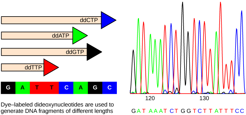

Figure 14.8図 14.8
In Frederick Sanger's dideoxy chain termination method, dye-labeled dideoxynucleotides are used to generate DNA fragments that terminate at different points. The DNA is separated by capillary electrophoresis on the basis of size, and from the order of fragments formed, the DNA sequence can be read. The DNA sequence readout is shown on an electropherogram that is generated by a laser scanner.
英語のヒント
フレデリック・サンガーのジデオキシ鎖終結法では、色素で標識したジデオキシヌクレオチドを用いて、それぞれ異なる位置で伸長が止まったDNA断片を作る。DNAをキャピラリー電気泳動によって長さに応じて分離し、できた断片の並びからDNA配列を読み取る。DNA配列の読取り結果は、レーザースキャナーで作成したエレクトロフェログラムに示される。
The DNA sample to be sequenced is denatured (separated into two strands by heating it to high temperatures). The DNA is divided into four tubes in which a primer, DNA polymerase, and all four nucleoside triphosphates (A, T, G, and C) are added. In addition, limited quantities of one of the four dideoxynucleoside triphosphates (ddCTP, ddATP, ddGTP, and ddTTP) are added to each tube respectively. The tubes are labeled as A, T, G, and C according to the ddNTP added. For detection purposes, each of the four dideoxynucleotides carries a different fluorescent label. Chain elongation continues until a fluorescent dideoxy nucleotide is incorporated, after which no further elongation takes place. After the reaction is over, electrophoresis is performed. Even a difference in length of a single base can be detected. The sequence is read from a laser scanner that detects the fluorescent marker of each fragment. For his work on DNA sequencing, Sanger received a Nobel Prize in Chemistry in 1980.
英語のヒント
配列を決定するDNA試料を変性させる(高温に加熱して2本の鎖に分ける)。DNAを4本の試験管に分け、それぞれにプライマー、DNAポリメラーゼ、4種類すべてのヌクレオシド三リン酸(A、T、G、C)を加える。さらに、4種類のジデオキシヌクレオシド三リン酸(ddCTP、ddATP、ddGTP、ddTTP)のうち1種類を、各試験管にそれぞれ少量ずつ加える。試験管には、加えたddNTPに応じてA、T、G、Cのラベルを付ける。検出のため、4種類のジデオキシヌクレオチドには、それぞれ異なる蛍光標識が付いている。鎖の伸長は、蛍光標識を付けたジデオキシヌクレオチドが取り込まれるまで続き、その後はそれ以上伸長しない。反応が終わったら電気泳動を行う。長さがわずか1塩基違うだけでも検出できる。各断片の蛍光標識を検出するレーザースキャナーから、配列を読み取る。サンガーはDNA配列決定の研究により、1980年にノーベル化学賞を受賞した。
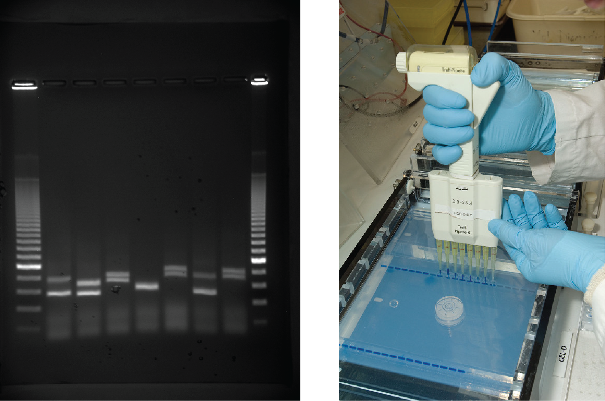
Gel electrophoresis is a technique used to separate DNA fragments of different sizes. Usually the gel is made of a chemical called agarose (a polysaccharide polymer extracted from seaweed that is high in galactose residues). Agarose powder is added to a buffer and heated. After cooling, the gel solution is poured into a casting tray. Once the gel has solidified, the DNA is loaded on the gel and electric current is applied. The DNA has a net negative charge and moves from the negative electrode toward the positive electrode. The electric current is applied for sufficient time to let the DNA separate according to size; the smallest fragments will be farthest from the well (where the DNA was loaded), and the heavier molecular weight fragments will be closest to the well. Once the DNA is separated, the gel is stained with a DNA-specific dye for viewing it (Figure 14.9).
英語のヒント
ゲル電気泳動は、長さの異なるDNA断片を分離する技術である。通常、ゲルにはアガロースという物質を用いる。これは、海藻から抽出される、ガラクトース残基に富む多糖の高分子である。アガロース粉末を緩衝液に加えて加熱する。冷却後、ゲル溶液を成型トレイに注ぐ。ゲルが固まったら、DNAをゲルに載せて電流を流す。DNAは全体として負の電荷をもち、負極から正極へ向かって移動する。長さに応じてDNAが分離するのに十分な時間、電流を流す。最も小さい断片はウェル(DNAを入れた場所)から最も遠くへ移動し、分子量の大きい断片はウェルの最も近くにとどまる。分離が済んだら、DNAに特異的な色素でゲルを染めて観察する(図14.9)。
OpenStax, Rice University; Biology 2e. (credit: James Jacob, Tompkins Cortland Community College) · CC BY 4.0 · Source出典

Figure 14.9図 14.9
DNA can be separated on the basis of size using gel electrophoresis. (credit: James Jacob, Tompkins Cortland Community College)
DNAは、ゲル電気泳動を用いて長さに応じて分離できる。(クレジット:James Jacob、トンプキンス・コートランド・コミュニティ・カレッジ)