第14章DNAの構造と機能
14.1 Historical Basis of Modern Understanding14.1 現在の理解の歴史的基盤
Learning Objectives学習目標
By the end of this section, you will be able to do the following:
この節を学ぶと、次のことができるようになる。
- Explain transformation of DNADNAによる形質転換を説明する。
- Describe the key experiments that helped identify that DNA is the genetic materialDNAが遺伝物質であると突き止めるうえで役立った、主要な実験を説明する。
- State and explain Chargaff’s rulesシャルガフの規則を述べ、説明する。
Our current understanding of DNA began with the discovery of nucleic acids followed by the development of the double-helix model. In the 1860s, Friedrich Miescher (Figure 14.2), a physician by profession, isolated phosphate-rich chemicals from white blood cells (leukocytes). He named these chemicals (which would eventually be known as DNA) nuclein because they were isolated from the nuclei of the cells.
英語のヒント
現在のDNAに関する理解は、核酸の発見に始まり、その後の二重らせんモデルの構築へとつながった。1860年代、医師を職業とするフリードリヒ・ミーシェル(図14.2)は、白血球からリン酸に富む化学物質を単離した。彼は、のちにDNAとして知られることになるこれらの物質を、細胞の核から単離したことにちなんでヌクレインと名づけた。
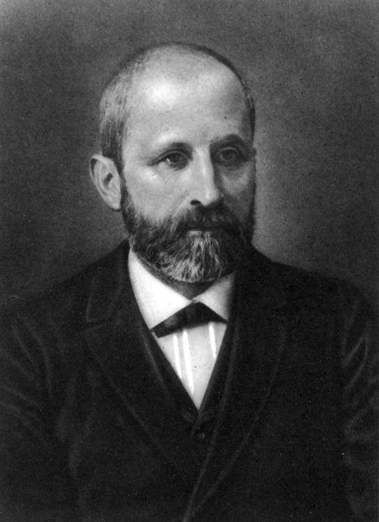
Figure 14.2図 14.2
Friedrich Miescher (1844–1895) discovered nucleic acids.
フリードリヒ・ミーシェル(1844〜1895年)は核酸を発見した。
A half century later, in 1928, British bacteriologist Frederick Griffith reported the first demonstration of bacterial transformation—a process in which external DNA is taken up by a cell, thereby changing its morphology and physiology. Griffith conducted his experiments with Streptococcus pneumoniae, a bacterium that causes pneumonia. Griffith worked with two strains of this bacterium called rough (R) and smooth (S). (The two cell types were called “rough” and “smooth” after the appearance of their colonies grown on a nutrient agar plate.)
英語のヒント
半世紀後の1928年、イギリスの細菌学者フレデリック・グリフィスは、細菌の形質転換を初めて実証したと報告した。形質転換とは、細胞が外部のDNAを取り込み、それによって形態や生理機能が変化する過程である。グリフィスは、肺炎を引き起こす細菌である肺炎球菌(Streptococcus pneumoniae)を使って実験した。彼が用いたのは、ラフ型(R型)とスムース型(S型)と呼ばれる、この細菌の2つの菌株だった。(この2種類の細胞は、栄養寒天培地上で増殖させたコロニーの外観にちなんで、「粗い」を意味するroughと「滑らかな」を意味するsmoothと呼ばれた。)
The R strain is non-pathogenic (does not cause disease). The S strain is pathogenic (disease-causing), and has a capsule outside its cell wall. The capsule allows the cell to escape the immune responses of the host mouse.
英語のヒント
R型菌は非病原性であり、病気を引き起こさない。S型菌は病原性、つまり病気を引き起こす性質があり、細胞壁の外側に莢膜をもつ。この莢膜によって、細胞は宿主であるマウスの免疫応答を免れることができる。
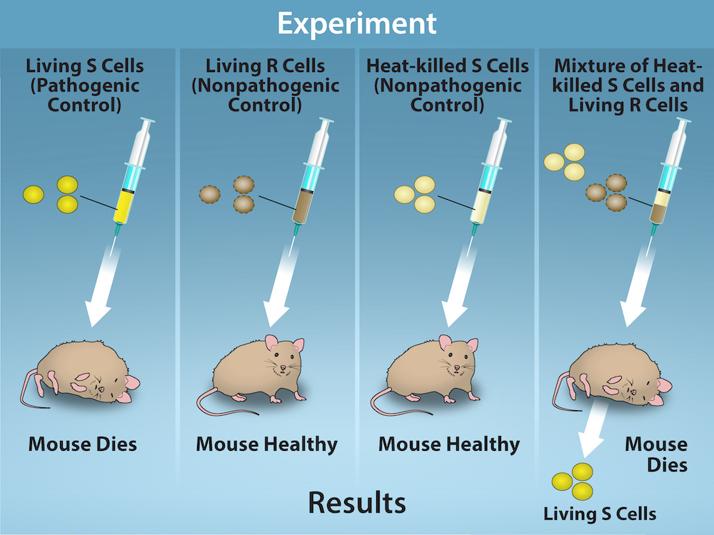
When Griffith injected the living S strain into mice, they died from pneumonia. In contrast, when Griffith injected the live R strain into mice, they survived. In another experiment, when he injected mice with the heat-killed S strain, they also survived. This experiment showed that the capsule alone was not the cause of death. In a third set of experiments, a mixture of live R strain and heat-killed S strain were injected into mice, and—to his surprise—the mice died. Upon isolating the live bacteria from the dead mouse, only the S strain of bacteria was recovered. When this isolated S strain was injected into fresh mice, the mice died. Griffith concluded that something had passed from the heat-killed S strain into the live R strain and transformed it into the pathogenic S strain. He called this the transforming principle (Figure 14.3). These experiments are now known as Griffith's transformation experiments.
英語のヒント
グリフィスが生きたS型菌をマウスに注射すると、マウスは肺炎で死んだ。これに対し、生きたR型菌を注射したマウスは生き残った。別の実験では、加熱して殺したS型菌を注射したマウスも生き残った。この実験から、莢膜だけが死の原因ではないことが示された。3番目の一連の実験では、生きたR型菌と加熱して殺したS型菌の混合物をマウスに注射した。すると、驚いたことにマウスは死んだ。死んだマウスから生きた細菌を単離すると、回収されたのはS型菌だけだった。この単離したS型菌を別のマウスに注射すると、そのマウスも死んだ。グリフィスは、加熱して殺したS型菌から生きたR型菌へ何かが移り、R型菌を病原性のS型菌に変化させたと結論づけた。彼はこれを形質転換因子と呼んだ(図14.3)。これらの実験は、現在ではグリフィスの形質転換実験として知られている。
OpenStax, Rice University; Biology 2e. Credit: Rao, A., Ryan, K. and Tag, A. Department of Biology, Texas A&M University. · CC BY 4.0 · Source出典
Figure 14.3図 14.3
Two strains of S. pneumoniae were used in Griffith’s transformation experiments. The R strain is non-pathogenic, whereas the S strain is pathogenic and causes death. When Griffith injected a mouse with the heat-killed S strain and a live R strain, the mouse died. The S strain was recovered from the dead mouse. Griffith concluded that something had passed from the heat-killed S strain to the R strain, transforming the R strain into the S strain in the process. Credit: Rao, A., Ryan, K. and Tag, A. Department of Biology, Texas A&M University.
英語のヒント
グリフィスの形質転換実験には、肺炎球菌(S. pneumoniae)の2つの菌株が用いられた。R型菌は非病原性だが、S型菌は病原性であり、死を引き起こす。グリフィスが、加熱して殺したS型菌と生きたR型菌をマウスに注射すると、マウスは死んだ。死んだマウスからはS型菌が回収された。グリフィスは、加熱して殺したS型菌からR型菌へ何かが移り、その過程でR型菌をS型菌に変えたと結論づけた。クレジット:Rao, A., Ryan, K. and Tag, A.、テキサスA&M大学生物学科。
Scientists Oswald Avery, Colin MacLeod, and Maclyn McCarty (1944) were interested in exploring this transforming principle further. They isolated the S strain from the dead mice and isolated the proteins and nucleic acids (RNA and DNA) as these were possible candidates for the molecule of heredity. They used enzymes that specifically degraded each component and then used each mixture separately to transform the R strain. They found that when DNA was degraded, the resulting mixture was no longer able to transform the bacteria, whereas all of the other combinations were able to transform the bacteria. This led them to conclude that DNA was the transforming principle.
英語のヒント
科学者のオズワルド・エイブリー、コリン・マクロード、マクリン・マッカーティ(1944年)は、この形質転換因子をさらに調べたいと考えた。彼らは死んだマウスからS型菌を単離し、さらにそのタンパク質と核酸(RNAとDNA)を単離した。これらが遺伝を担う分子の候補だったためである。彼らは、それぞれの成分を特異的に分解する酵素を使い、処理後の各混合物を別々に用いてR型菌を形質転換させた。その結果、DNAを分解した場合には、得られた混合物はもはや細菌を形質転換できなかったが、ほかの組み合わせではすべて形質転換が起こることがわかった。こうして彼らは、DNAが形質転換因子であると結論づけた。
Although the experiments of Avery, McCarty and McLeod had demonstrated that DNA was the informational component transferred during transformation, DNA was still considered to be too simple a molecule to carry biological information. Proteins, with their 20 different amino acids, were regarded as more likely candidates. The decisive experiment, conducted by Martha Chase and Alfred Hershey in 1952, provided confirmatory evidence that DNA was indeed the genetic material and not proteins. Chase and Hershey were studying a bacteriophage—a virus that infects bacteria. Viruses typically have a simple structure: a protein coat, called the capsid, and a nucleic acid core that contains the genetic material (either DNA or RNA). The bacteriophage infects the host bacterial cell by attaching to its surface, and then it injects its nucleic acids inside the cell. The phage DNA makes multiple copies of itself using the host machinery, and eventually the host cell bursts, releasing a large number of bacteriophages. Hershey and Chase selected radioactive elements that would specifically distinguish the protein from the DNA in infected cells. They labeled one batch of phage with radioactive sulfur, 35S, to label the protein coat. Another batch of phage were labeled with radioactive phosphorus, 32P. Because phosphorous is found in DNA, but not protein, the DNA and not the protein would be tagged with radioactive phosphorus. Likewise, sulfur is absent from DNA, but present in several amino acids such as methionine and cysteine.
英語のヒント
エイブリー、マッカーティ、マクロードの実験によって、形質転換の際に移される情報の担い手がDNAであることが示されていた。それでもDNAは、生物学的情報を担うには単純すぎる分子だと考えられていた。20種類のアミノ酸からなるタンパク質のほうが、有力な候補とみなされていた。1952年にマーサ・チェイスとアルフレッド・ハーシーが行った決定的な実験は、遺伝物質がタンパク質ではなくDNAであることを裏づける証拠を示した。チェイスとハーシーが研究していたのは、細菌に感染するウイルスであるバクテリオファージだった。ウイルスは通常、カプシドと呼ばれるタンパク質の殻と、遺伝物質(DNAまたはRNA)を含む核酸の芯という、単純な構造をもつ。バクテリオファージは、宿主となる細菌の表面に付着して感染し、続いて核酸を細胞内へ注入する。ファージのDNAは宿主の仕組みを利用して自分自身のコピーを多数作り、やがて宿主細胞が破裂して、多数のバクテリオファージを放出する。ハーシーとチェイスは、感染細胞の中でタンパク質とDNAを特異的に区別できる放射性元素を選んだ。一群のファージでは、放射性硫黄35Sを使ってタンパク質の殻を標識した。別の一群のファージは、放射性リン32Pで標識した。リンはDNAには含まれるがタンパク質には含まれないため、放射性リンで標識されるのはタンパク質ではなくDNAとなる。同様に、硫黄はDNAには含まれないが、メチオニンやシステインなど、いくつかのアミノ酸に含まれる。
Each batch of phage was allowed to infect the cells separately. After infection, the phage bacterial suspension was put in a blender, which caused the phage coat to detach from the host cell. Cells exposed long enough for infection to occur were then examined to see which of the two radioactive molecules had entered the cell. The phage and bacterial suspension was spun down in a centrifuge. The heavier bacterial cells settled down and formed a pellet, whereas the lighter phage particles stayed in the supernatant. In the tube that contained phage labeled with 35S, the supernatant contained the radioactively labeled phage, whereas no radioactivity was detected in the pellet. In the tube that contained the phage labeled with 32P, the radioactivity was detected in the pellet that contained the heavier bacterial cells, and no radioactivity was detected in the supernatant. Hershey and Chase concluded that it was the phage DNA that was injected into the cell and carried information to produce more phage particles, thus providing evidence that DNA was the genetic material and not proteins (Figure 14.4).
英語のヒント
それぞれの一群のファージを別々に細胞に感染させた。感染後、ファージと細菌の懸濁液をミキサーにかけ、ファージの殻を宿主細胞から引き離した。感染が起こるのに十分な時間ファージにさらした細胞を調べ、2種類の放射性分子のうちどちらが細胞内に入ったかを確認した。ファージと細菌の懸濁液を遠心分離した。重い細菌の細胞は沈んでペレットを形成し、軽いファージ粒子は上清に残った。 35Sで標識したファージを含む試験管では、放射性標識を付けたファージが上清に含まれ、ペレットからは放射能が検出されなかった。32Pで標識したファージを含む試験管では、重い細菌の細胞を含むペレットから放射能が検出され、上清からは検出されなかった。ハーシーとチェイスは、細胞に注入され、より多くのファージ粒子を作るための情報を運んだのはファージのDNAであると結論づけた。こうして、遺伝物質がタンパク質ではなくDNAである証拠が示された(図14.4)。
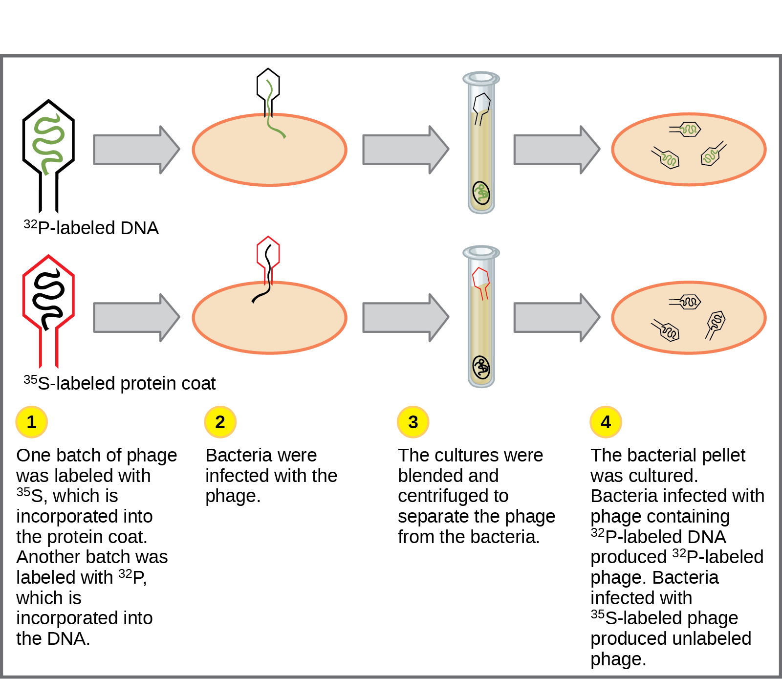


Figure 14.4図 14.4
In Hershey and Chase's experiments, bacteria were infected with phage radiolabeled with either 35S, which labels protein, or 32P, which labels DNA. Only 32P entered the bacterial cells, indicating that DNA is the genetic material.
英語のヒント
ハーシーとチェイスの実験では、タンパク質を標識する35S、またはDNAを標識する32Pのいずれかで放射性標識を付けたファージを、細菌に感染させた。細菌の細胞内に入ったのは32Pだけであり、DNAが遺伝物質であることを示した。
Around this same time, Austrian biochemist Erwin Chargaff examined the content of DNA in different species and found that the amounts of adenine, thymine, guanine, and cytosine were not found in equal quantities, and that relative concentrations of the four nucleotide bases varied from species to species, but not within tissues of the same individual or between individuals of the same species. He also discovered something unexpected: That the amount of adenine equaled the amount of thymine, and the amount of cytosine equaled the amount of guanine (that is, A = T and G = C). Different species had equal amounts of purines (A+G) and pyrimidines (T + C), but different ratios of A+T to G+C. These observations became known as Chargaff’s rules. Chargaff's findings proved immensely useful when Watson and Crick were getting ready to propose their DNA double helix model! You can see after reading the past few pages how science builds upon previous discoveries, sometimes in a slow and laborious process.
英語のヒント
ほぼ同じころ、オーストリアの生化学者エルヴィン・シャルガフは、さまざまな種のDNAの組成を調べた。その結果、アデニン、チミン、グアニン、シトシンは同じ量ずつ存在するわけではなく、4種類のヌクレオチド塩基の相対的な濃度は種によって異なることを見いだした。しかし、同じ個体の組織間や、同じ種の個体間では異ならなかった。彼は、予想外のことも発見した。アデニンの量はチミンの量に等しく、シトシンの量はグアニンの量に等しかったのである(つまり、A = T、G = C)。異なる種でも、プリン塩基(A + G)とピリミジン塩基(T + C)の量は等しかったが、A + TとG + Cの比は異なっていた。これらの観察結果は、シャルガフの規則として知られるようになった。ワトソンとクリックがDNA二重らせんモデルを提案する準備をしていたとき、シャルガフの発見はきわめて役に立った。ここまでの数ページを読めば、科学が過去の発見を土台にして築かれ、その過程が時にはゆっくりとした骨の折れるものになることがわかるだろう。