第14章DNAの構造と機能
14.2 DNA Structure and Sequencing14.2 DNAの構造と配列決定
Learning Objectives学習目標
By the end of this section, you will be able to do the following:
この節を学ぶと、次のことができるようになる。
- Describe the structure of DNADNAの構造を説明する。
- Explain the Sanger method of DNA sequencingDNA配列決定のサンガー法を説明する。
- Discuss the similarities and differences between eukaryotic and prokaryotic DNA真核生物と原核生物のDNAの共通点と相違点について論じる。
The building blocks of DNA are nucleotides. The important components of the nucleotide are a nitrogenous (nitrogen-bearing) base, a 5-carbon sugar (pentose), and a phosphate group (Figure 14.5). The nucleotide is named depending on the nitrogenous base. The nitrogenous base can be a purine such as adenine (A) and guanine (G), or a pyrimidine such as cytosine (C) and thymine (T).
英語のヒント
DNAの基本的な構成単位はヌクレオチドである。ヌクレオチドの重要な構成成分は、含窒素塩基、炭素原子5個をもつ糖(五炭糖)、リン酸基である(図14.5)。ヌクレオチドの名前は含窒素塩基に応じて付けられる。含窒素塩基には、アデニン(A)やグアニン(G)のようなプリン塩基と、シトシン(C)やチミン(T)のようなピリミジン塩基がある。
The purines have a double ring structure with a six-membered ring fused to a five-membered ring. Pyrimidines are smaller in size; they have a single six-membered ring structure.
プリン塩基は、六員環と五員環が融合した二環構造をもつ。ピリミジン塩基はそれより小さく、1つの六員環構造をもつ。
The sugar is deoxyribose in DNA and ribose in RNA. The carbon atoms of the five-carbon sugar are numbered 1', 2', 3', 4', and 5' (1' is read as “one prime”). The phosphate, which makes DNA and RNA acidic, is connected to the 5' carbon of the sugar by the formation of an ester linkage between phosphoric acid and the 5'-OH group (an ester is an acid + an alcohol). In DNA nucleotides, the 3' carbon of the sugar deoxyribose is attached to a hydroxyl (OH) group. In RNA nucleotides, the 2' carbon of the sugar ribose also contains a hydroxyl group. The base is attached to the 1'carbon of the sugar.
英語のヒント
糖は、DNAではデオキシリボース、RNAではリボースである。五炭糖の炭素原子には、1′、2′、3′、4′、5′という番号が付いている(1′は「ワン・プライム」と読む)。DNAとRNAを酸性にするリン酸は、リン酸と5′-OH基との間にエステル結合ができることによって、糖の5′炭素に結びつく(エステルは酸とアルコールからできる)。DNAのヌクレオチドでは、糖であるデオキシリボースの3′炭素にヒドロキシ基(OH)が結合している。RNAのヌクレオチドでは、糖であるリボースの2′炭素にもヒドロキシ基がある。塩基は糖の1′炭素に結合している。
The nucleotides combine with each other to produce phosphodiester bonds. The phosphate residue attached to the 5' carbon of the sugar of one nucleotide forms a second ester linkage with the hydroxyl group of the 3' carbon of the sugar of the next nucleotide, thereby forming a 5'-3' phosphodiester bond. In a polynucleotide, one end of the chain has a free 5' phosphate, and the other end has a free 3'-OH. These are called the 5' and 3' ends of the chain. A base attached to a five-carbon sugar represents a nucleoside. A nucleotide is composed of one, two, or three phosphate groups attached to a nucleoside.
英語のヒント
ヌクレオチドどうしは、ホスホジエステル結合を作ってつながる。あるヌクレオチドの糖の5′炭素に付いたリン酸残基が、次のヌクレオチドの糖の3′炭素にあるヒドロキシ基と2つ目のエステル結合を作る。こうして5′–3′ホスホジエステル結合が形成される。ポリヌクレオチドでは、鎖の一端に遊離の5′リン酸基があり、もう一端に遊離の3′-OHがある。これらは鎖の5′末端、3′末端と呼ばれる。塩基が五炭糖に結合したものがヌクレオシドである。ヌクレオチドは、ヌクレオシドに1個、2個、または3個のリン酸基が結合してできている。
In the 1950s, Francis Crick and James Watson worked together to determine the structure of DNA at the University of Cambridge, England. Other scientists like Linus Pauling and Maurice Wilkins were also actively exploring this field. Pauling previously had discovered the secondary structure of proteins using X-ray crystallography. In Wilkins’ lab, researcher Rosalind Franklin was using X-ray diffraction methods to understand the structure of DNA. Watson and Crick were able to piece together the puzzle of the DNA molecule on the basis of Franklin's data because Crick had also studied X-ray diffraction (Figure 14.6). In 1962, James Watson, Francis Crick, and Maurice Wilkins were awarded the Nobel Prize in Medicine. Unfortunately, by then Franklin had died, and Nobel prizes are not awarded posthumously.
英語のヒント
1950年代、フランシス・クリックとジェームズ・ワトソンは、イギリスのケンブリッジ大学でDNAの構造を解明するために共同研究を行った。ライナス・ポーリングやモーリス・ウィルキンスなど、ほかの科学者もこの分野を精力的に研究していた。ポーリングはそれ以前に、X線結晶構造解析を使ってタンパク質の二次構造を発見していた。ウィルキンスの研究室では、研究者のロザリンド・フランクリンが、DNAの構造を理解するためにX線回折法を用いていた。クリックもX線回折を研究していたため、ワトソンとクリックはフランクリンのデータに基づいて、DNA分子というパズルを組み立てることができた(図14.6)。1962年、ジェームズ・ワトソン、フランシス・クリック、モーリス・ウィルキンスはノーベル医学生理学賞を受賞した。残念ながら、その時点ですでにフランクリンは亡くなっており、ノーベル賞は死後に授与されることはない。
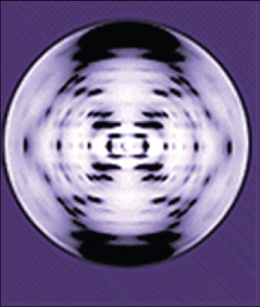
Figure 14.6図 14.6
The X-ray diffraction pattern of DNA, which helped to elucidate its double-helix structure.
英語のヒント
DNAの二重らせん構造の解明に役立った、DNAのX線回折像。
Watson and Crick proposed that DNA is made up of two strands that are twisted around each other to form a right-handed helix. Base pairing takes place between a purine and pyrimidine on opposite strands, so that A pairs with T, and G pairs with C (suggested by Chargaff's Rules). Thus, adenine and thymine are complementary base pairs, and cytosine and guanine are also complementary base pairs. The base pairs are stabilized by hydrogen bonds: adenine and thymine form two hydrogen bonds and cytosine and guanine form three hydrogen bonds. The two strands are anti-parallel in nature; that is, the 3' end of one strand faces the 5' end of the other strand. The sugar and phosphate of the nucleotides form the backbone of the structure, whereas the nitrogenous bases are stacked inside, like the rungs of a ladder. Each base pair is separated from the next base pair by a distance of 0.34 nm, and each turn of the helix measures 3.4 nm. Therefore, 10 base pairs are present per turn of the helix. The diameter of the DNA double-helix is 2 nm, and it is uniform throughout. Only the pairing between a purine and pyrimidine and the antiparallel orientation of the two DNA strands can explain the uniform diameter. The twisting of the two strands around each other results in the formation of uniformly spaced major and minor grooves (Figure 14.7).
英語のヒント
ワトソンとクリックは、DNAが互いに巻きつき、右巻きのらせんを作る2本の鎖からなると提案した。塩基対は、向かい合う鎖のプリン塩基とピリミジン塩基の間で形成され、AはTと、GはCと対を作る(シャルガフの規則によって示唆された)。したがって、アデニンとチミンは相補的な塩基対であり、シトシンとグアニンも相補的な塩基対である。塩基対は水素結合によって安定化される。アデニンとチミンは2本、シトシンとグアニンは3本の水素結合を形成する。2本の鎖は逆平行である。つまり、一方の鎖の3′末端が、もう一方の鎖の5′末端に向かい合っている。ヌクレオチドの糖とリン酸が構造の骨格を形成し、含窒素塩基ははしごの横木のように内側に積み重なる。各塩基対と次の塩基対との距離は0.34 nmであり、らせん1回転の長さは3.4 nmである。したがって、らせん1回転あたり10塩基対が存在する。DNA二重らせんの直径は2 nmで、全体を通じて一定である。プリン塩基とピリミジン塩基の対形成と、2本のDNA鎖が逆平行に並ぶことだけが、この一定の直径を説明できる。2本の鎖が互いに巻きつくことで、主溝と副溝が等間隔に形成される(図14.7)。
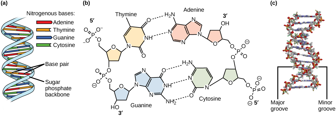


Figure 14.7図 14.7
DNA has (a) a double helix structure and (b) phosphodiester bonds; the dotted lines between Thymine and Adenine and Guanine and Cytosine represent hydrogen bonds. The (c) major and minor grooves are binding sites for DNA binding proteins during processes such as transcription (the copying of RNA from DNA) and replication.
英語のヒント
DNAには、(a)二重らせん構造と(b)ホスホジエステル結合がある。チミンとアデニンの間、およびグアニンとシトシンの間の点線は、水素結合を表す。(c)主溝と副溝は、転写(DNAからRNAをコピーすること)や複製などの過程で、DNA結合タンパク質の結合部位となる。