13.2 Chromosomal Basis of Inherited Disorders13.2 遺伝性疾患の染色体上の基盤
Learning Objectives学習目標
By the end of this section, you will be able to do the following:
この節を学ぶと、次のことができるようになる。
Describe how a karyogram is created
核型図の作り方を説明する。
Explain how nondisjunction leads to disorders in chromosome number
不分離が染色体数の異常を生むしくみを説明する。
Compare disorders that aneuploidy causes
異数性による疾患を比較する。
Describe how errors in chromosome structure occur through inversions and translocations
逆位と転座を通じて染色体構造の誤りが生じるしくみを説明する。
Inherited disorders can arise when chromosomes behave abnormally during meiosis. We can divide chromosome disorders into two categories: abnormalities in chromosome number and chromosomal structural rearrangements. Because even small chromosome segments can span many genes, chromosomal disorders are characteristically dramatic and often fatal.
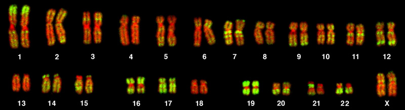
Chromosome isolation and microscopic observation forms the basis of cytogenetics and is the primary method by which clinicians detect chromosomal abnormalities in humans. A karyotype is the number and appearance of chromosomes, and includes their length, banding pattern, and centromere position. To obtain a view of an individual’s karyotype, cytologists photograph the chromosomes and then cut and paste each chromosome into a chart, or karyogram. Another name is an ideogram (Figure 13.5).
OpenStax, Rice University; Biology 2e. (credit: Andreas Blozer et al) · CC BY 4.0 · Source出典
Figure 13.5図 13.5
This karyotype is of a female human. Notice that homologous chromosomes are the same size, and have the same centromere positions and banding patterns. A human male would have an XY chromosome pair instead of the XX pair. (credit: Andreas Blozer et al)
In a given species, we can identify chromosomes by their number, size, centromere position, and banding pattern. In a human karyotype, autosomes or “body chromosomes” (all of the non–sex chromosomes) are generally organized in approximate order of size from largest (chromosome 1) to smallest (chromosome 22). The X and Y chromosomes are not autosomes. However, chromosome 21 is actually shorter than chromosome 22. Researchers discovered this after naming Down syndrome as trisomy 21, reflecting how this disorder results from possessing one extra chromosome 21 (three total). Not wanting to change the name of this important disorder, scientists retained the numbering of chromosome 21 despite describing it having the shortest set of chromosomes. We may designate the chromosome “arms” projecting from either end of the centromere as short or long, depending on their relative lengths. We abbreviate the short arm p (for “petite”); whereas, we abbreviate the long arm q (because it follows “p” alphabetically). Numbers further subdivide and denote each arm. Using this naming system, we can describe chromosome locations consistently in the scientific literature.