第11章減数分裂と有性生殖
Anaphase I第一分裂後期
In anaphase I, the microtubules pull the linked chromosomes apart. The sister chromatids remain tightly bound together at the centromere. The chiasmata are broken in anaphase I as the microtubules attached to the fused kinetochores pull the homologous chromosomes apart (Figure 11.6).
第一分裂後期には微小管が、つながった染色体どうしを引き離す。姉妹染色分体はセントロメアで強くつながったままである。結合した動原体に付着する微小管が相同染色体を引き離すにつれ、キアズマが解消される(図11.6)。
Telophase I and Cytokinesis第一分裂終期と細胞質分裂
In telophase, the separated chromosomes arrive at opposite poles. The remainder of the typical telophase events may or may not occur, depending on the species. In some organisms, the chromosomes “decondense” and nuclear envelopes form around the separated sets of chromatids produced during telophase I. In other organisms, cytokinesis—the physical separation of the cytoplasmic components into two daughter cells—occurs without reformation of the nuclei. In nearly all species of animals and some fungi, cytokinesis separates the cell contents via a cleavage furrow (constriction of the actin ring that leads to cytoplasmic division). In plants, a cell plate is formed during cell cytokinesis by Golgi vesicles fusing at the metaphase plate. This cell plate will ultimately lead to the formation of cell walls that separate the two daughter cells.
英語のヒント
終期には分離した染色体が反対側の極へ到着する。それ以外の典型的な終期の出来事は、種によって起こる場合も起こらない場合もある。染色体の凝縮がほどけ、第一分裂終期にできた別々の染色分体の組を核膜が包む生物もいれば、核が再形成されないまま、細胞質の成分を2個の娘細胞へ物理的に分ける細胞質分裂が起こる生物もいる。動物のほぼ全種と一部の菌類では、分裂溝によって細胞内容物を分ける。これはアクチン環が収縮して細胞質を分割するものである。植物では、細胞質分裂中にゴルジ小胞が赤道板で融合して細胞板を作る。これが最終的に2個の娘細胞を隔てる細胞壁の形成につながる。
Two haploid cells are the result of the first meiotic division of a diploid cell. The cells are haploid because at each pole, there is just one of each pair of the homologous chromosomes. Therefore, only one full set of the chromosomes is present. This is why the cells are considered haploid—there is only one chromosome set, even though each chromosome still consists of two sister chromatids. Recall that sister chromatids are merely duplicates of one of the two homologous chromosomes (except for changes that occurred during crossing over). In meiosis II, these two sister chromatids will separate, creating four haploid daughter cells.
英語のヒント
二倍体細胞の減数第一分裂の結果、2個の半数体細胞ができる。各極には相同染色体の各対の片方しかなく、完全な染色体の組は1組だけになるので半数体である。各染色体がまだ2本の姉妹染色分体からなっていても、組が1組しかないため半数体と見なす。姉妹染色分体は、乗換えで起こった変化を除けば、2本の相同染色体の片方の複製にすぎない。減数第二分裂でこの2本が分離し、4個の半数体の娘細胞ができる。
Meiosis II減数第二分裂
In some species, cells enter a brief interphase, or interkinesis, before entering meiosis II. Interkinesis lacks an S phase, so chromosomes are not duplicated. The two cells produced in meiosis I go through the events of meiosis II in synchrony. During meiosis II, the sister chromatids within the two daughter cells separate, forming four new haploid gametes. The mechanics of meiosis II are similar to mitosis, except that each dividing cell has only one set of homologous chromosomes, each with two chromatids. Therefore, each cell has half the number of sister chromatids to separate out as a diploid cell undergoing mitosis. In terms of chromosomal content, cells at the start of meiosis II are similar to haploid cells in G2, preparing to undergo mitosis.
英語のヒント
一部の種では、減数第二分裂の前に、分裂間期という短い間期に入る。分裂間期にはS期がないため、染色体は複製されない。第一分裂でできた2細胞は、第二分裂を同調して進める。二つの娘細胞内の姉妹染色分体が分離し、4個の新しい半数体配偶子ができる。第二分裂のしくみは有糸分裂に似ているが、分裂する各細胞は、各2本の染色分体からなる相同染色体を1組だけ持つ。そのため分離する姉妹染色分体の数は、有糸分裂する二倍体細胞の半分である。染色体の内容から見ると、第二分裂の開始時の細胞は、有糸分裂を準備するG2期の半数体細胞に似ている。
Prophase II第二分裂前期
If the chromosomes decondensed in telophase I, they condense again. If nuclear envelopes were formed, they fragment into vesicles. The MTOCs that were duplicated during interkinesis move away from each other toward opposite poles, and new spindles are formed.
英語のヒント
第一分裂終期に染色体の凝縮がほどけていた場合は、再び凝縮する。核膜が形成されていた場合は、小胞へと断片化する。分裂間期に複製された微小管形成中心(MTOC)は、互いに離れて反対側の極へ移動し、新しい紡錘体が形成される。
Prometaphase II第二分裂前中期
The nuclear envelopes are completely broken down, and the spindle is fully formed. Each sister chromatid forms an individual kinetochore that attaches to microtubules from opposite poles.
英語のヒント
核膜は完全に分解され、紡錘体が完成する。それぞれの姉妹染色分体に個別の動原体ができ、反対側の極から伸びた微小管が付着する。
Metaphase II第二分裂中期
The sister chromatids are maximally condensed and aligned at the equator of the cell.
英語のヒント
姉妹染色分体は最大限に凝縮し、細胞の赤道面に並ぶ。
Anaphase II第二分裂後期
The sister chromatids are pulled apart by the kinetochore microtubules and move toward opposite poles. Nonkinetochore microtubules elongate the cell.
姉妹染色分体は動原体微小管によって引き離され、反対側の極へ移動する。非動原体微小管は細胞を伸長させる。
OpenStax, Rice University; Biology 2e. Credit: Rao, A., Ryan, K., Fletcher, S. and Tag, A. Department of Biology, Texas A&M University. · CC BY 4.0 · Source出典
Figure 11.6図 11.6
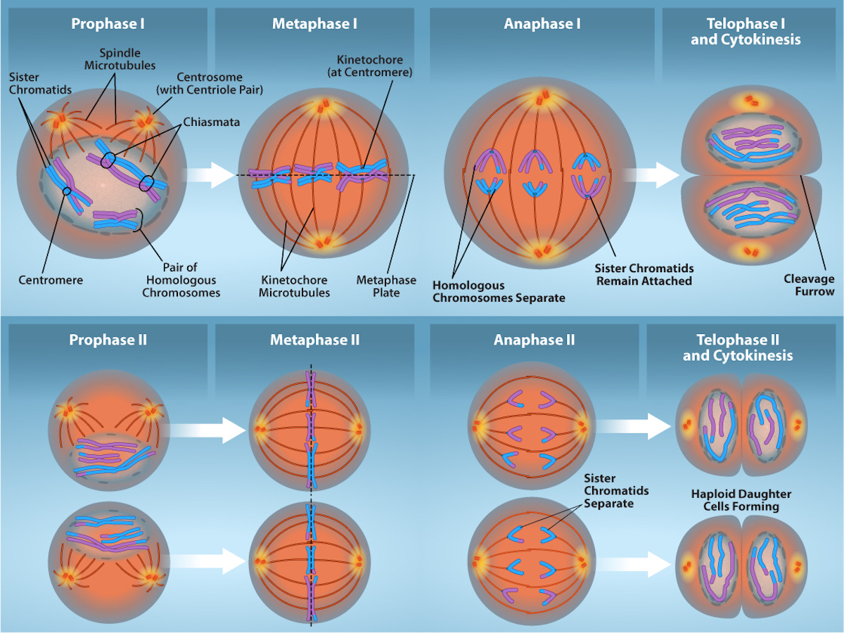
The process of chromosome alignment differs between meiosis I and meiosis II. In prophase I, pairs of homologous chromosomes form chiasmata which allow for crossing over events (genetic diversity). These pairs of homologous chromosomes arrange at the metaphase plate in metaphase I. In anaphase I, homologous chromosomes separate. Telophase I and cytokinesis result in haploid cells with 2 sister chromatids of each chromosome. In prophase II, spindle microtubules form and elongate and any nuclear envelope disappears. Sister chromatids arrange at the midpoint of the cells in metaphase II. In anaphase II, the sister chromatids separate. Telophase II and cytokinesis result in haploid cells with a single copy of each chromosome. Credit: Rao, A., Ryan, K., Fletcher, S. and Tag, A. Department of Biology, Texas A&M University.
染色体が並ぶ過程は減数第一分裂と第二分裂で異なる。第一分裂前期に相同染色体の対がキアズマを形成し、乗換えが可能になる(遺伝的多様性)。この相同染色体対が第一分裂中期に赤道板に並ぶ。第一分裂後期に相同染色体が分離する。第一分裂終期と細胞質分裂の結果、各染色体が2本の姉妹染色分体からなる半数体細胞ができる。第二分裂前期に紡錘体微小管が形成されて伸長し、核膜があれば消失する。第二分裂中期に姉妹染色分体が細胞の中央に並ぶ。第二分裂後期に姉妹染色分体が分離する。第二分裂終期と細胞質分裂の結果、各染色体を1本ずつ持つ半数体細胞ができる。出典:Rao, A.、Ryan, K.、Fletcher, S.、Tag, A.、テキサスA&M大学生物学科。
Telophase II and Cytokinesis第二分裂終期と細胞質分裂
The chromosomes arrive at opposite poles and begin to decondense. Nuclear envelopes form around the chromosomes. If the parent cell was diploid, as is most commonly the case, then cytokinesis now separates the two cells into four unique haploid cells. The cells produced are genetically unique because of the random assortment of paternal and maternal homologs and because of the recombination of maternal and paternal segments of chromosomes (with their sets of genes) that occurs during crossover. The entire process of meiosis is outlined in Figure 11.7.
英語のヒント
染色体は反対側の極に到着し、凝縮がほどけ始める。染色体を囲む核膜ができる。最も普通に見られるように親細胞が二倍体だった場合、ここで細胞質分裂が2個の細胞を分け、遺伝的に異なる4個の半数体細胞を作る。できた細胞が遺伝的に固有なのは、父親由来と母親由来の相同染色体が無作為に振り分けられること、そして乗換えで母親由来と父親由来の染色体部分が、その部分の遺伝子群ごと組み換えられることによる。図11.7に減数分裂の全過程を示す。
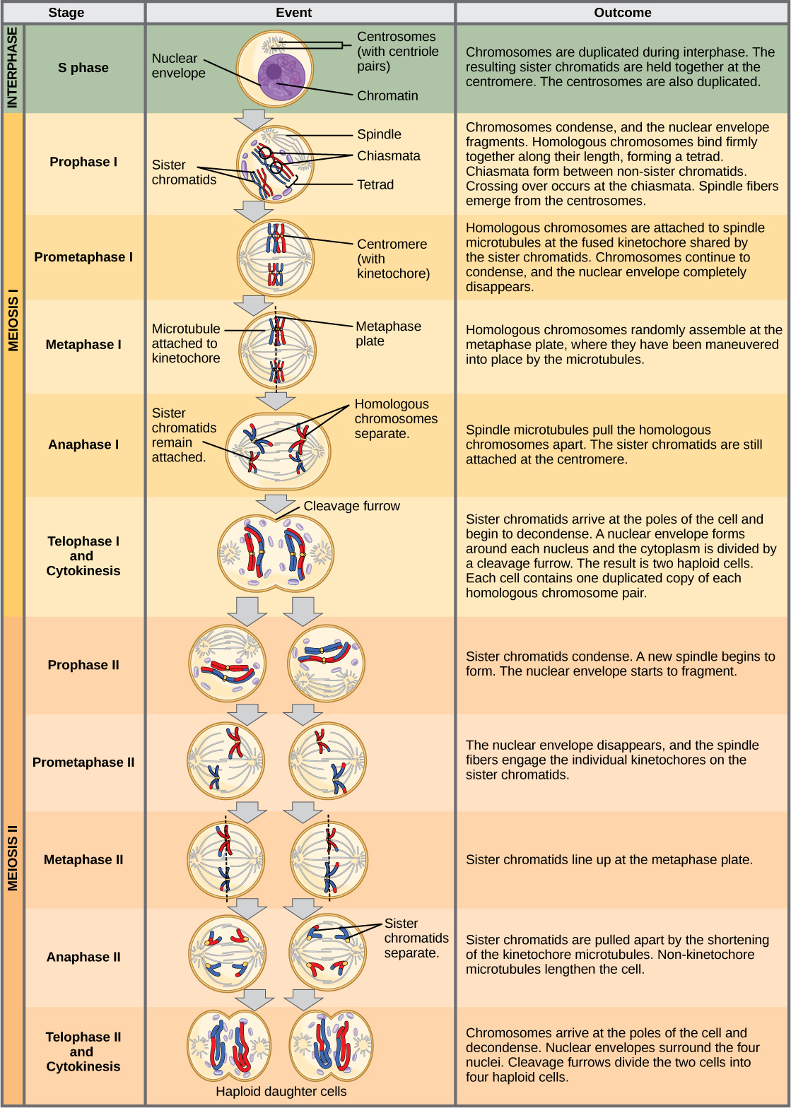


Figure 11.7図 11.7
An animal cell with a diploid number of four (2n = 4) proceeds through the stages of meiosis to form four haploid daughter cells.
二倍体の染色体数が4本(2n=4)の動物細胞は、減数分裂の各段階を経て、4個の半数体の娘細胞を形成する。