第22章原子The Atom原書 p.743–794
22.5 Medical Applications of Radioactivity: Diagnostic Imaging and Radiation22.5 放射能の医療への応用——画像診断と放射線治療
SECTION LEARNING OBJECTIVES By the end of this section, you will be able to do the following:
この節の学習目標 この節を終えると、次のことができるようになる。
• Describe how nuclear imaging works (e.g., radioisotope imaging, PET)• Describe the ionizing effects of radiation and how they can be used for medical treatment
・核医学の画像(放射性同位体による画像、PETなど)がどうはたらくかを述べる・放射線の電離の効果と、それが医療の治療にどう使えるかを述べる
Section Key Termsこの節の重要語
Anger cameraradradiopharmaceuticaltherapeutic ratiorelative biological effectiveness (RBE)roentgen equivalent man (rem)tagged
治療の アンガーカメラ ラド 放射性医薬品 比生物学的効果比(RBE)レム(roentgen equivalent man) 標識された
Medical Applications of Nuclear Physics核物理の医療への応用
Applications of nuclear physics have become an integral part of modern life. From the bone scan that detects one cancer to the radioiodine treatment that cures another, nuclear radiation has diagnostic and therapeutic effects on medicine.
核物理の応用は現代の生活に欠かせないものになった。あるがんを見つける骨シンチグラフィーから、別のがんを治す放射性ヨウ素の治療まで、核の放射線は医療で診断と治療の両方の効果を持つ。
Medical Imaging A host of medical imaging techniques employ nuclear radiation. What makes nuclear radiation so useful? First,
医療の画像 多くの医療の画像の技術が核の放射線を使う。核の放射線がこれほど役に立つのはなぜか。第一に、
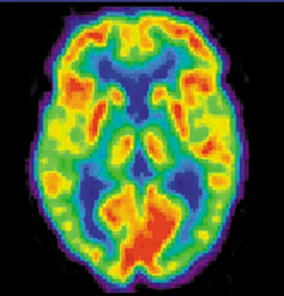
radiation can easily penetrate tissue; hence, it is a useful probe to monitor conditions inside the body. Second, nuclear radiation depends on the nuclide and not on the chemical compound it is in, so that a radioactive nuclide can be put into a compound designed for specific purposes. When that is done, the compound is said to be tagged. A tagged compound used for medical purposes is called a radiopharmaceutical. Radiation detectors external to the body can determine the location and concentration of a radiopharmaceutical to yield medically useful information. For example, certain drugs are concentrated in inflamed regions of the body, and their locations can aid diagnosis and treatment as seen in Figure 22.37. Another application utilizes a radiopharmaceutical that the body sends to bone cells, particularly those that are most active, to detect cancerous tumors or healing points. Images can then be produced of such bone scans. Clever use of radioisotopes determines the functioning of body organs, such as blood flow, heart muscle activity, and iodine uptake in the thyroid gland. For instance, a radioactive form of iodine can be used to monitor the thyroid, a radioactive thallium salt can be used to follow the blood stream, and radioactive gallium can be used for cancer imaging.
放射線は組織を容易に突き抜ける。したがって体の中の様子を調べる有用な探りになる。第二に、核の放射線はどの核種かによって決まり、それがどんな化合物の中にあるかにはよらない。だから放射性の核種を、特定の目的に合わせて作った化合物に組み込める。そうしたとき、その化合物は標識されたという。医療の目的で使われる標識された化合物を放射性医薬品という。体の外の放射線の検出器が放射性医薬品の位置と濃さを定め、医療に役立つ情報を与える。たとえばある種の薬は体の炎症のある場所に濃く集まり、その位置が図22.37のように診断と治療の助けになる。別の応用では、体が骨の細胞、とくに最も活発な細胞へ送る放射性医薬品を使い、がんの腫瘍や治りかけの箇所を見つける。そうした骨のシンチグラフィーの画像を作れる。放射性同位体の巧みな使い方によって、血の流れ、心筋のはたらき、甲状腺のヨウ素の取り込みなど、体の器官のはたらきが定められる。たとえば放射性の形のヨウ素は甲状腺を調べるのに、放射性のタリウムの塩は血の流れを追うのに、放射性のガリウムはがんの画像に使える。
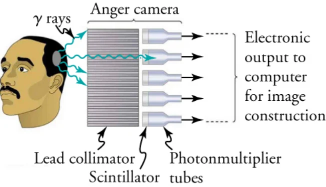
Once a radioactive compound has been ingested, a device like that shown in Figure 22.38 is used to monitor nuclear activity. The device, called an Anger camera or gamma camera uses a piece of lead with holes bored through it. The gamma rays are redirected through the collimator to narrow their beam, and are then interpreted using a device called a scintillator. The computer analysis of detector signals produces an image. One of the disadvantages of this detection method is that there is no depth information (i.e., it provides a two-dimensional view of the tumor as opposed to a three-dimensional view), because radiation from any location under that detector produces a signal.
放射性の化合物を取り込ませたあと、図22.38のような装置で核の活動を調べる。アンガーカメラ、あるいはガンマカメラと呼ばれるこの装置は、穴をあけた鉛の板を使う。ガンマ線はコリメータを通されて束が細く絞られ、それからシンチレータという装置で読み取られる。検出器の信号をコンピュータが分析して画像を作る。この検出法の弱点の一つは、深さの情報がないことである(つまり腫瘍を三次元ではなく二次元で見ることになる)。検出器の下のどの位置からの放射線でも信号を作ってしまうからである。
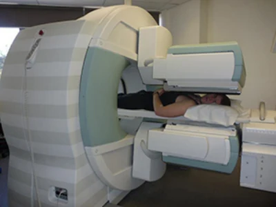
Single-photon-emission computer tomography (SPECT) used in conjunction with a CT scanner improves on the process carried out by the gamma camera. Figure 22.39 shows a patient in a circular array of SPECT detectors that may be stationary or rotated, with detector output used by a computer to construct a detailed image. The spatial resolution of this technique is poor, but the three-dimensional image created results in a marked improvement in contrast.
CT装置と組み合わせて使う単一光子放射断層撮影(SPECT)は、ガンマカメラの行う過程を改良したものである。図22.39は、止まったまま、あるいは回るSPECTの検出器の輪の中にいる患者を示す。検出器の出力をコンピュータが使って細かい画像を組み立てる。この手法は空間の分解能は低いが、三次元の画像が作られるため、濃淡の違いが著しく改善される。
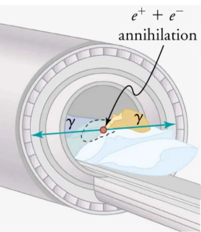
Positron emission tomography (or PET) scans utilize images produced by emitters. When the emitted positron β+ encounters an electron, mutual annihilation occurs, producing two γ rays. Those rays have identical 0.511 MeV energies (the energy comes from the destruction of an electron or positron mass) and they move directly away from each other, allowing detectors to determine their point of origin accurately (as shown in Figure 22.40). It requires detectors on opposite sides to simultaneously (i.e., at the same time) detect photons of 0.511 MeV energy and utilizes computer imaging techniques similar to those in SPECT and CT scans. PET is used extensively for diagnosing brain disorders. It can note decreased metabolism in certain regions that accompany Alzheimer’s disease. PET can also locate regions in the brain that become active when a person carries out specific activities, such as speaking, closing his or her eyes, and so on.
陽電子放出断層撮影(PET)は、を放つ核種が作る画像を使う。放たれた陽電子β+が電子と出会うと対消滅が起こり、二つのγ線ができる。この線はどちらも0.511 MeVの同じエネルギーを持ち(このエネルギーは電子または陽電子の質量が消えることから来る)、互いにまっすぐ反対の向きへ進むので、検出器はその出どころを正確に定められる(図22.40に示す)。反対側どうしの検出器が0.511 MeVのエネルギーの光子を同時に(つまり同じ時刻に)検出する必要があり、SPECTやCTと似たコンピュータの画像化の技術を使う。PETは脳の疾患の診断に広く使われる。アルツハイマー病に伴う、ある領域の代謝の低下を見てとれる。PETはまた、話す、目を閉じるといった特定の行いをするときに活発になる脳の領域を突き止めることもできる。
Ionizing Radiation on the Body体に対する電離放射線
We hear many seemingly contradictory things about the biological effects of ionizing radiation. It can cause cancer, burns, and hair loss, and yet it is used to treat and even cure cancer. How do we understand such effects? Once again, there is an underlying simplicity in nature, even in complicated biological organisms. All the effects of ionizing radiation on biological tissue can be understood by knowing that ionizing radiation affects molecules within cells, particularly DNA molecules. Let us take a brief look at molecules within cells and how cells operate. Cells have long, double-helical DNA molecules containing chemical patterns called genetic codes that govern the function and processes undertaken by the cells. Damage to DNA consists of breaks in chemical bonds or other changes in the structural features of the DNA chain, leading to changes in the genetic code. In human cells, we can have as many as a million individual instances of damage to DNA per cell per day. The repair ability of DNA is vital for maintaining the integrity of the genetic code and for the normal functioning of the entire organism. A cell with a damaged ability to repair DNA, which could have been induced by ionizing radiation, can do one of the following:
電離放射線の生物への効果について、私たちは一見矛盾する話をいろいろ聞く。がんや火傷や脱毛を起こしうるのに、がんの治療、さらには治癒にも使われる。こうした効果をどう理解すればよいのか。ここでもまた、込み入った生き物であっても、自然の底には単純さがある。電離放射線が生物の組織に及ぼす効果はすべて、電離放射線が細胞の中の分子、とくにDNAの分子に作用すると知れば理解できる。細胞の中の分子と、細胞がどうはたらくかを手短に見よう。細胞は長い二重らせんのDNAの分子を持ち、そこには遺伝の暗号と呼ばれる化学の模様が収められていて、細胞のはたらきと営みを支配している。DNAの損傷とは、化学結合が切れることや、DNAの鎖の構造上の特徴が変わることであり、遺伝の暗号の変化につながる。人の細胞では、細胞一つあたり一日に100万件ものDNAの損傷が起こりうる。DNAの修復の力は、遺伝の暗号の完全さを保ち、体全体が正しくはたらくために欠かせない。電離放射線によって起こされたかもしれないDNAの修復の力の損なわれた細胞は、次のいずれかになりうる。
• The cell can go into an irreversible state of dormancy, known as senescence.• The cell can commit suicide, known as programmed cell death.• The cell can go into unregulated cell division, leading to tumors and cancers.
・細胞は、老化と呼ばれる後戻りできない眠りの状態に入りうる。・細胞は、計画された細胞の死と呼ばれる自殺をしうる。・細胞は、調節を失った細胞分裂に入り、腫瘍やがんにつながりうる。
Since ionizing radiation damages the DNA, ionizing radiation has its greatest effect on cells that rapidly reproduce, including most types of cancer. Thus, cancer cells are more sensitive to radiation than normal cells and can be killed by it easily. Cancer is characterized by a malfunction of cell reproduction, and can also be caused by ionizing radiation. There is no contradiction to say that ionizing radiation can be both a cure and a cause.
電離放射線はDNAを傷つけるので、電離放射線は速く増える細胞に最も強く効く。ほとんどの型のがんがそれにあたる。したがってがんの細胞はふつうの細胞より放射線に敏感で、放射線で容易に殺せる。がんは細胞の増殖の狂いを特徴とし、電離放射線によって起こされることもある。電離放射線が治療にもなり原因にもなると言っても、矛盾はない。
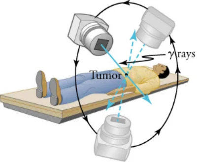
Radiotherapy Radiotherapy is effective against cancer because cancer cells reproduce rapidly and, consequently, are more sensitive to radiation. The central problem in radiotherapy is to make the dose for cancer cells as high as possible while limiting the dose for normal cells. The ratio of abnormal cells killed to normal cells killed is called the therapeutic ratio, and all radiotherapy techniques are designed to enhance that ratio. Radiation can be concentrated in cancerous tissue by a number of techniques. One of the most prevalent techniques for well-defined tumors is a geometric technique shown in Figure 22.41. A narrow beam of radiation is passed through the patient from a variety of directions with a common crossing point in the tumor. The technique concentrates the dose in the tumor while spreading it out over a large volume of normal tissue.
放射線治療 放射線治療ががんに効くのは、がんの細胞が速く増え、その結果として放射線に敏感だからである。放射線治療の中心の課題は、ふつうの細胞への線量を抑えつつ、がんの細胞への線量をできるだけ大きくすることである。殺される異常な細胞と殺されるふつうの細胞の比を治療比といい、放射線治療の技術はすべてこの比を高めるように作られている。放射線をがんの組織に集める技術はいくつもある。境目のはっきりした腫瘍に最も広く使われる技術の一つが、図22.41に示す幾何学的な技術である。細く絞った放射線の束を、腫瘍で交わる一点を共有するように、いろいろな向きから患者に通す。この技術は線量を腫瘍に集めつつ、ふつうの組織には大きな体積にわたって薄く散らす。
Another use of radiation therapy is through radiopharmaceuticals. Cleverly, radiopharmaceuticals are used in cancer therapy by tagging antibodies with radioisotopes. Those antibodies are extracted from the patient, cultured, loaded with a radioisotope, and then returned to the patient. The antibodies are then concentrated almost entirely in the tissue they developed to fight, thus localizing the radiation in abnormal tissue. This method is used with radioactive iodine to fight thyroid cancer. While the therapeutic ratio can be quite high for such short-range radiation, there can be a significant dose for organs that eliminate radiopharmaceuticals from the body, such as the liver, kidneys, and bladder. As with most radiotherapy, the technique is limited by the tolerable amount of damage to the normal tissue.
放射線治療のもう一つの使い方が放射性医薬品である。巧みなことに、放射性医薬品は抗体を放射性同位体で標識することでがんの治療に使われる。抗体は患者から取り出され、培養され、放射性同位体を積まれ、それから患者に戻される。抗体はそれが戦うために育った組織にほぼすべて集まるので、放射線が異常な組織に集まることになる。この方法は放射性ヨウ素を使って甲状腺がんと戦うのに用いられる。そうした届く範囲の短い放射線では治療比をかなり高くできるが、肝臓・腎臓・膀胱など、放射性医薬品を体から取り除く器官にはかなりの線量が及びうる。多くの放射線治療と同じく、この技術はふつうの組織が耐えられる損傷の量で頭打ちになる。
Radiation Dosage To quantitatively discuss the biological effects of ionizing radiation, we need a radiation dose unit that is directly related to those effects. To do define such a unit, it is important to consider both the biological organism and the radiation itself. Knowing that the amount of ionization is proportional to the amount of deposited energy, we define a radiation dose unit called the rad. It 1/100 of a joule of ionizing energy deposited per kilogram of tissue, which is
放射線の線量 電離放射線の生物への効果を量として論じるには、その効果に直接結びつく線量の単位が要る。そうした単位を定めるには、生き物と放射線そのものの両方を考えることが大切である。電離の量は与えられたエネルギーの量に比例するので、ラドという放射線の線量の単位を定める。それは組織1キログラムあたりに与えられる電離のエネルギーの100分の1ジュールであり、
For example, if a 50.0-kg person is exposed to ionizing radiation over her entire body and she absorbs 1.00 J, then her whole-body radiation dose is
たとえば体重50.0 kgの人が全身に電離放射線を浴び、1.00 Jを吸収したとすると、その人の全身の放射線の線量は
If the same 1.00 J of ionizing energy were absorbed in her 2.00-kg forearm alone, then the dose to the forearm would be
同じ1.00 Jの電離のエネルギーが、2.00 kgの前腕だけに吸収されたとすると、前腕への線量は
and the unaffected tissue would have a zero rad dose. When calculating radiation doses, you divide the energy absorbed by the mass of affected tissue. You must specify the affected region, such as the whole body or forearm in addition to giving the numerical dose in rads. Although the energy per kilogram in 1 rad is small, it can still have significant effects. Since only a few eV cause ionization, just 0.01 J of ionizing energy can create a huge number of ion pairs and have an effect at the cellular level.
となり、影響を受けていない組織の線量は0ラドになる。放射線の線量を計算するときは、吸収されたエネルギーを影響を受けた組織の質量で割る。ラドで表した数値だけでなく、全身なのか前腕なのかといった影響を受けた領域も示さなければならない。1ラドの1キログラムあたりのエネルギーは小さいが、それでも大きな効果を持ちうる。電離を起こすのに要るのはわずか数eVなので、たった0.01 Jの電離のエネルギーでもぼう大な数のイオン対を作り、細胞の水準で効果を及ぼしうる。
The effects of ionizing radiation may be directly proportional to the dose in rads, but they also depend on the type of radiation and the type of tissue. That is, for a given dose in rads, the effects depend on whether the radiation is , , , X-ray, or some other type of ionizing radiation. The relative biological effectiveness (RBE) relates to the amount of biological damage that can occur from a given type of radiation and is given in Table 22.4 for several types of ionizing radiation.
電離放射線の効果はラドで表した線量に正比例しうるが、放射線の型と組織の型にもよる。つまり、ラドで表した同じ線量でも、効果はその放射線がかかかX線か、あるいは別の型の電離放射線かによる。生物学的効果比(RBE)は、ある型の放射線が起こしうる生物の損傷の量に関わり、いくつかの型の電離放射線について表22.4に示す。
TIPS FOR SUCCESS The RBEs given in Table 22.4 are approximate, but they yield certain valuable insights.
学習のポイント 表22.4のRBEはおおよその値だが、いくつか価値ある見通しを与えてくれる。
• The eyes are more sensitive to radiation, because the cells of the lens do not repair themselves.• Though both are neutral and have large ranges, neutrons cause more damage than rays because neutrons often cause secondary radiation when they are captured.• Short-range particles such as rays have a severely damaging effect to internal anatomy, as their damage is concentrated and more difficult for the biological organism to repair. However, the skin can usually block alpha particles from entering the body.
・目は放射線により敏感である。水晶体の細胞は自分を修復しないからである。・中性子も線もどちらも中性で届く範囲が長いが、中性子のほうが損傷が大きい。中性子は捕らえられるときにしばしば二次の放射線を作るからである。・線のような届く範囲の短い粒子は、体の内部に激しい損傷を与える。損傷が一点に集まり、生き物にとって修復がより難しいからである。ただし皮膚はふつうアルファ粒子が体内へ入るのを止められる。
Can you think of any other insights from the table?
表から他にどんな見通しが読み取れるだろうか。
A final dose unit more closely related to the effect of radiation on biological tissue is called the roentgen equivalent man, or rem. A combination of all factors mentioned previously, the roentgen equivalent man is defined to be the dose in rads multiplied by the relative biological effectiveness.
放射線の生物の組織への効果により近く結びついた最後の線量の単位を、レム(roentgen equivalent man)という。これまでに挙げたすべての要素を合わせたもので、レムはラドで表した線量に生物学的効果比を掛けたものと定められる。
The large-scale effects of radiation on humans can be divided into two categories: immediate effects and long-term effects. Table 22.5 gives the immediate effects of whole-body exposures received in less than one day. If the radiation exposure is spread out over more time, greater doses are needed to cause the effects listed. Any dose less than 10 rem is called a low dose, a dose 10 to 100 rem is called a moderate dose, and anything greater than 100 rem is called a high dose.
放射線が人に及ぼす大きな効果は、すぐ現れる効果と長い時間をかけて現れる効果の二つに分けられる。表22.5は、一日足らずで全身に浴びた場合のすぐ現れる効果を示す。同じ被曝がもっと長い時間に散らばれば、挙げられた効果を起こすにはより大きな線量が要る。10レム未満の線量を低線量、10から100レムを中線量、100レムを超えるものを高線量という。
Health Physicist Are you interested in learning more about radiation? Are you curious about studying radiation dosage levels and ensuring the safety of the environment and people that are most closely affected by it? If so, you may be interested in becoming a health physicist.
保健物理士 放射線についてもっと学びたいだろうか。放射線の線量を調べ、環境と、それに最も近く関わる人々の安全を守ることに興味があるだろうか。それなら保健物理士になることに関心を持つかもしれない。
The field of health physics draws from a variety of science disciplines with the central aim of mitigating radiation concerns. Those that work as health physicists have a diverse array of potential jobs available to them, including those in research, industry, education, environmental protection, and governmental regulation. Furthermore, while the term health physicist may lead many to think of the medical field, there are plenty of applications within the military, industrial, and energy fields as well.
保健物理という分野は、放射線の懸念を和らげることを中心のねらいとして、さまざまな科学の分野から材料を引いてくる。保健物理士として働く人には、研究・産業・教育・環境保護・行政の規制など、多様な仕事の可能性が開かれている。さらに、保健物理士という言葉から医療の分野を思い浮かべる人が多いだろうが、軍事・産業・エネルギーの分野にも応用は数多くある。
As a researcher, a health physicist can further environmental studies on the effects of radiation, design instruments for more accurate measurements, and assist in establishing valuable radiation standards. Within the energy field, a health physicsist often acts as a manager, closely tied to all operations at all levels, from procuring appropirate equipment to monitoring health data. Within industry, the health physicist acts as a consultant, assisting industry management in important decisions, designing facilities, and choosing appropriate detection tools. The health physicist possesses a unique knowledge base that allows him or her to operate in a wide variety of interesting disciplines!
研究者としての保健物理士は、放射線の効果についての環境の研究を進め、より正確に測る装置を設計し、価値ある放射線の基準を定めるのを助けられる。エネルギーの分野では、保健物理士はしばしば管理者として、適切な装置の調達から健康データの監視まで、あらゆる水準のすべての運用に密に関わる。産業では、保健物理士は助言者として、産業の経営陣の重要な判断を助け、施設を設計し、適切な検出の道具を選ぶ。保健物理士は独特の知識の土台を持ち、それによって非常に幅広い面白い分野で仕事ができるのである。
To become a health physicist, it is necessary to have a background in the physical sciences. Understanding the fields of biology, physiology, biochemistry, and genetics are all important as well. The ability to analyze and solve new problems is critical, and a natural aptitude for science and mathematics will assist in the continued necessary training. There are two possible certifications for health physicists: from the American Board of Health Physicists (ABHP) and the National Registry of Radiation Protection Technologists (NRRPT).
保健物理士になるには、自然科学の素養が要る。生物学・生理学・生化学・遺伝学の分野を理解することもみな大切である。新しい問題を分析して解く力は欠かせず、科学と数学への生まれつきの適性は、その後も必要になる訓練を助ける。保健物理士には二つの資格がある。米国保健物理士認定機構(ABHP)と、放射線防護技術者全国登録(NRRPT)のものである。