第5章感覚と知覚
5.3 Vision5.3 視覚
Learning Objectives学習目標
By the end of this section, you will be able to:
この節を学び終えると、次のことができるようになる。
- Describe the basic anatomy of the visual system視覚系の基本的な解剖学的構造を説明する
- Discuss how rods and cones contribute to different aspects of vision桿体と錐体が視覚のさまざまな側面にどのように寄与しているかを論じる
- Describe how monocular and binocular cues are used in the perception of depth奥行きの知覚において、単眼手がかりと両眼手がかりがどのように使われるかを説明する
The visual system constructs a mental representation of the world around us (Figure 5.10). This contributes to our ability to successfully navigate through physical space and interact with important individuals and objects in our environments. This section will provide an overview of the basic anatomy and function of the visual system. In addition, we will explore our ability to perceive color and depth.
英語のヒント
視覚系は、私たちを取り巻く世界の心的表象を組み立てる(図5.10)。このことは、物理的な空間の中をうまく動き回り、環境の中の重要な人や物とかかわる私たちの能力に寄与している。この節では、視覚系の基本的な解剖学的構造と機能を概観する。さらに、色と奥行きを知覚する私たちの能力を探っていく。

OpenStax, Rice University; Psychology 2e. (credit "top left”: modification of work by "rajkumar1220"/Flickr"; credit “top right”: modification of work by Thomas Leuthard; credit “middle left”: modification of work by Demietrich Baker; credit “middle right”: modification of work by "kaybee07"/Flickr; credit “bottom left”: modification of work by "Isengardt"/Flickr; credit “bottom right”: modification of work by Willem Heerbaart) · CC BY 4.0 · Source出典
Figure 5.10図 5.10
Our eyes take in sensory information that helps us understand the world around us. (credit "top left”: modification of work by "rajkumar1220"/Flickr"; credit “top right”: modification of work by Thomas Leuthard; credit “middle left”: modification of work by Demietrich Baker; credit “middle right”: modification of work by "kaybee07"/Flickr; credit “bottom left”: modification of work by "Isengardt"/Flickr; credit “bottom right”: modification of work by Willem Heerbaart)
私たちの目は、身の回りの世界を理解するのに役立つ感覚情報を取り込む。(出典「上段左」:“rajkumar1220”/Flickrの作品を改変、出典「上段右」:Thomas Leuthardの作品を改変、出典「中段左」:Demietrich Bakerの作品を改変、出典「中段右」:“kaybee07”/Flickrの作品を改変、出典「下段左」:“Isengardt”/Flickrの作品を改変、出典「下段右」:Willem Heerbaartの作品を改変)
Anatomy of the Visual System視覚系の解剖学的構造
The eye is the major sensory organ involved in vision (Figure 5.11). Light waves are transmitted across the cornea and enter the eye through the pupil. The cornea is the transparent covering over the eye. It serves as a barrier between the inner eye and the outside world, and it is involved in focusing light waves that enter the eye. The pupil is the small opening in the eye through which light passes, and the size of the pupil can change as a function of light levels as well as emotional arousal. When light levels are low, the pupil will become dilated, or expanded, to allow more light to enter the eye. When light levels are high, the pupil will constrict, or become smaller, to reduce the amount of light that enters the eye. The pupil’s size is controlled by muscles that are connected to the iris, which is the colored portion of the eye.
英語のヒント
目は、視覚にかかわる主要な感覚器官である(図5.11)。光波は角膜を通過し、瞳孔から目の中に入る。角膜は、目の表面を覆う透明な膜である。角膜は目の内部と外界とを隔てる障壁として働き、また、目に入ってくる光波の焦点を合わせることにもかかわっている。瞳孔は、光が通り抜ける目の小さな開口部であり、瞳孔の大きさは、光の量や感情的な覚醒に応じて変化しうる。光の量が少ないときには、より多くの光を目に入れるために、瞳孔は散大する、つまり広がる。光の量が多いときには、目に入る光の量を減らすために、瞳孔は収縮する、つまり小さくなる。瞳孔の大きさは、目の色のついた部分である虹彩につながった筋肉によって調節される。
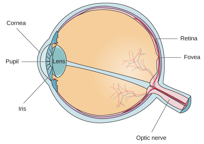
Figure 5.11図 5.11
The anatomy of the eye is illustrated in this diagram.
この図は、目の解剖学的構造を示している。
After passing through the pupil, light crosses the lens, a curved, transparent structure that serves to provide additional focus. The lens is attached to muscles that can change its shape to aid in focusing light that is reflected from near or far objects. In a normal-sighted individual, the lens will focus images perfectly on a small indentation in the back of the eye known as the fovea, which is part of the retina, the light-sensitive lining of the eye. The fovea contains densely packed specialized photoreceptor cells (Figure 5.12). These photoreceptor cells, known as cones, are light-detecting cells. The cones are specialized types of photoreceptors that work best in bright light conditions. Cones are very sensitive to acute detail and provide tremendous spatial resolution. They are also directly involved in our ability to perceive color.
英語のヒント
瞳孔を通過した光は、次に水晶体を通る。水晶体は湾曲した透明な構造で、焦点をさらに合わせる働きをする。水晶体には、その形を変えられる筋肉がつながっており、これによって、近くや遠くの物体から反射してくる光の焦点を合わせるのを助けている。正常な視覚を持つ人では、水晶体は像を、目の奥にある中心窩と呼ばれる小さなくぼみに、ぴったりと結ぶ。中心窩は網膜、すなわち目の内側を覆う光に敏感な膜の一部である。中心窩には、特殊化した視細胞が密集している(図5.12)。錐体と呼ばれるこれらの視細胞は、光を検出する細胞である。錐体は、明るい光の条件で最もよく働く特殊な種類の視細胞である。錐体は細部に非常に敏感で、きわめて高い空間分解能をもたらす。また、色を知覚する私たちの能力にも直接かかわっている。
While cones are concentrated in the fovea, where images tend to be focused, rods, another type of photoreceptor, are located throughout the remainder of the retina. Rods are specialized photoreceptors that work well in low light conditions, and while they lack the spatial resolution and color function of the cones, they are involved in our vision in dimly lit environments as well as in our perception of movement on the periphery of our visual field.
英語のヒント
錐体が、像の焦点が結ばれることの多い中心窩に集中しているのに対し、もう一つの種類の視細胞である桿体は、網膜のそれ以外の部分全体に分布している。桿体は、光の少ない条件でよく働く特殊化した視細胞である。桿体は錐体のような空間分解能や色の機能を持たないが、薄暗い環境での視覚や、視野の周辺部での動きの知覚にかかわっている。
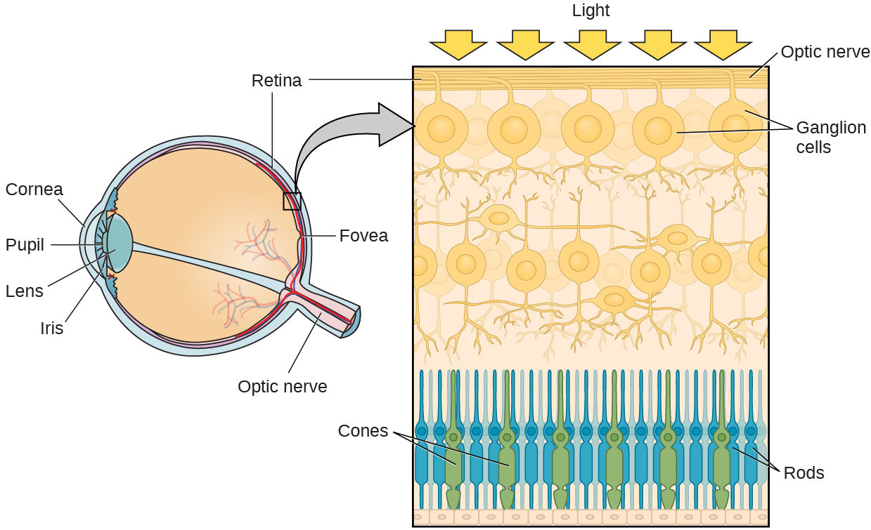
Figure 5.12図 5.12
The two types of photoreceptors are shown in this image. Cones are colored green and rods are blue.
この図には、2種類の視細胞が示されている。錐体は緑色、桿体は青色で描かれている。
Most of us have experienced the different sensitivities of rods and cones when making the transition from a brightly lit environment to a dimly lit environment. Imagine going to see a blockbuster movie on a clear summer day. As you walk from the brightly lit lobby into the dark theater, you notice that you immediately have difficulty seeing much of anything. After a few minutes, you begin to adjust to the darkness and can see the interior of the theater. In the bright environment, your vision was dominated primarily by cone activity. As you move to the dark environment, rod activity dominates, but there is a delay in transitioning between the phases. If your rods do not transform light into nerve impulses as easily and efficiently as they should, you will have difficulty seeing in dim light, a condition known as night blindness.
英語のヒント
私たちの多くは、明るく照らされた環境から薄暗い環境へ移るときに、桿体と錐体の感度の違いを経験したことがある。晴れた夏の日に、話題の大作映画を見に行くところを想像してみよう。明るいロビーから暗い劇場の中へ歩いていくと、入ってすぐは、ほとんど何もよく見えないことに気づく。数分たつと暗さに慣れはじめ、劇場の中が見えるようになる。明るい環境では、あなたの視覚は主に錐体の活動に支配されていた。暗い環境に移ると桿体の活動が優位になるが、二つの段階のあいだの移行には遅れがある。もし桿体が本来あるべきほど容易かつ効率的に光を神経インパルスに変換しないと、薄暗い光の中で見るのに苦労することになる。これは夜盲症として知られる状態である。
Rods and cones are connected (via several interneurons) to retinal ganglion cells. Axons from the retinal ganglion cells converge and exit through the back of the eye to form the optic nerve. The optic nerve carries visual information from the retina to the brain. There is a point in the visual field called the blind spot: Even when light from a small object is focused on the blind spot, we do not see it. We are not consciously aware of our blind spots for two reasons: First, each eye gets a slightly different view of the visual field; therefore, the blind spots do not overlap. Second, our visual system fills in the blind spot so that although we cannot respond to visual information that occurs in that portion of the visual field, we are also not aware that information is missing.
英語のヒント
桿体と錐体は(いくつかの介在ニューロンを介して)網膜神経節細胞につながっている。網膜神経節細胞から出た軸索は一つに集まり、目の奥から外へ出て視神経を形づくる。視神経は、視覚情報を網膜から脳へ運ぶ。視野の中には、盲点と呼ばれる点がある。小さな物体からの光が盲点に焦点を結んでも、私たちにはその物体が見えない。私たちが自分の盲点を意識しないのには、二つの理由がある。第一に、それぞれの目がとらえる視野はわずかに異なっているので、盲点どうしが重ならない。第二に、視覚系が盲点を埋めるため、視野のその部分に生じる視覚情報には反応できないにもかかわらず、情報が欠けていることにも気づかない。
The optic nerve from each eye merges just below the brain at a point called the optic chiasm. As Figure 5.13 shows, the optic chiasm is an X-shaped structure that sits just below the cerebral cortex at the front of the brain. At the point of the optic chiasm, information from the right visual field (which comes from both eyes) is sent to the left side of the brain, and information from the left visual field is sent to the right side of the brain.
英語のヒント
それぞれの目から出た視神経は、脳のすぐ下の、視交叉と呼ばれる地点で合流する。図5.13が示すように、視交叉はX字形の構造で、脳の前部で大脳皮質のすぐ下に位置している。視交叉の地点で、右視野からの情報(両目から来る)は脳の左側に送られ、左視野からの情報は脳の右側に送られる。
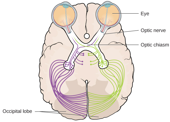


Figure 5.13図 5.13
This illustration shows the optic chiasm at the front of the brain and the pathways to the occipital lobe at the back of the brain, where visual sensations are processed into meaningful perceptions.
この図は、脳の前部にある視交叉と、脳の後部にある後頭葉へ向かう経路を示している。後頭葉では、視覚的な感覚が処理されて意味のある知覚になる。
Once inside the brain, visual information is sent via a number of structures to the occipital lobe at the back of the brain for processing. Visual information might be processed in parallel pathways which can generally be described as the “what pathway” and the “where/how” pathway. The “what pathway” is involved in object recognition and identification, while the “where/how pathway” is involved with location in space and how one might interact with a particular visual stimulus (Milner & Goodale, 2008; Ungerleider & Haxby, 1994). For example, when you see a ball rolling down the street, the “what pathway” identifies what the object is, and the “where/how pathway” identifies its location or movement in space.
英語のヒント
脳の中に入ると、視覚情報はいくつもの構造を経由して、処理のために脳の後部にある後頭葉へ送られる。視覚情報は並列する経路で処理されているのかもしれず、それらの経路はおおまかに「何」経路と「どこ/どのように」経路と表現できる。「何」経路は物体の認識と同定にかかわり、「どこ/どのように」経路は、空間内の位置と、特定の視覚刺激に対して人がどう働きかけうるかにかかわっている(Milner & Goodale, 2008; Ungerleider & Haxby, 1994)。たとえば、ボールが通りを転がっていくのを見たとき、「何」経路はその物体が何であるかを識別し、「どこ/どのように」経路はその物体の空間内での位置や動きを識別する。