第3章生物心理学
The Two Hemispheres二つの半球
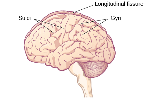
The surface of the brain, known as the cerebral cortex, is very uneven, characterized by a distinctive pattern of folds or bumps, known as gyri (singular: gyrus), and grooves, known as sulci (singular: sulcus), shown in Figure 3.15. These gyri and sulci form important landmarks that allow us to separate the brain into functional centers. The most prominent sulcus, known as the longitudinal fissure, is the deep groove that separates the brain into two halves or hemispheres: the left hemisphere and the right hemisphere.
英語のヒント
大脳皮質と呼ばれる脳の表面は非常に凹凸が多く、特徴的な模様をなしている。その模様は、脳回(gyri、単数形はgyrus)と呼ばれるひだや隆起と、脳溝(sulci、単数形はsulcus)と呼ばれる溝とから成る(図3.15)。これらの脳回と脳溝は重要な目印となり、それによって脳を機能ごとの中枢に区分できる。最も目立つ脳溝は大脳縦裂と呼ばれ、脳を二つの半分、すなわち二つの半球(左半球と右半球)に分ける深い溝である。
OpenStax, Rice University; Psychology 2e. (credit: modification of work by Bruce Blaus) · CC BY 4.0 · Source出典

Figure 3.15図 3.15
The surface of the brain is covered with gyri and sulci. A deep sulcus is called a fissure, such as the longitudinal fissure that divides the brain into left and right hemispheres. (credit: modification of work by Bruce Blaus)
脳の表面は脳回と脳溝に覆われている。深い脳溝は「裂」と呼ばれ、脳を左右の半球に分ける大脳縦裂はその一例である。(出典:Bruce Blausの作品を改変)
There is evidence of specialization of function—referred to as lateralization—in each hemisphere, mainly regarding differences in language functions. The left hemisphere controls the right half of the body, and the right hemisphere controls the left half of the body. Decades of research on lateralization of function by Michael Gazzaniga and his colleagues suggest that a variety of functions ranging from cause-and-effect reasoning to self-recognition may follow patterns that suggest some degree of hemispheric dominance (Gazzaniga, 2005). For example, the left hemisphere has been shown to be superior for forming associations in memory, selective attention, and positive emotions. The right hemisphere, on the other hand, has been shown to be superior in pitch perception, arousal, and negative emotions (Ehret, 2006). However, it should be pointed out that research on which hemisphere is dominant in a variety of different behaviors has produced inconsistent results, and therefore, it is probably better to think of how the two hemispheres interact to produce a given behavior rather than attributing certain behaviors to one hemisphere versus the other (Banich & Heller, 1998).
英語のヒント
それぞれの半球には、側性化と呼ばれる機能の特殊化が見られるという証拠があり、それは主に言語機能の違いにかかわるものである。左半球は体の右半分を制御し、右半球は体の左半分を制御している。マイケル・ガザニガと共同研究者たちによる数十年にわたる機能の側性化の研究は、因果推論から自己認識に至るまでのさまざまな機能が、ある程度の半球優位性をうかがわせるパターンに従っている可能性を示唆している(Gazzaniga, 2005)。たとえば、左半球は、記憶における連合の形成、選択的注意、ポジティブな感情において優れていることが示されている。一方、右半球は、音の高さの知覚、覚醒、ネガティブな感情において優れていることが示されている(Ehret, 2006)。しかし、さまざまな行動においてどちらの半球が優位なのかについての研究は、一貫しない結果を生んできたことを指摘しておくべきである。したがって、特定の行動を一方の半球か他方の半球かに帰属させるよりも、二つの半球がどのように相互作用してある行動を生み出しているのかを考える方がおそらくよい(Banich & Heller, 1998)。
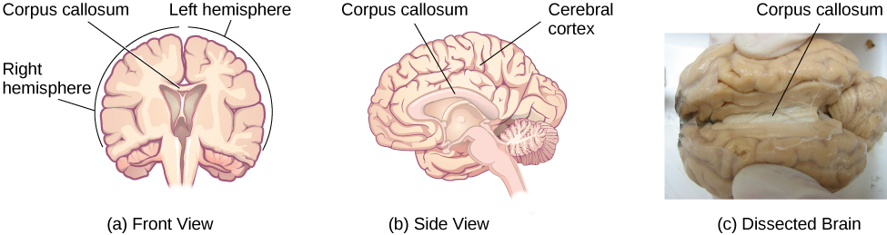
The two hemispheres are connected by a thick band of neural fibers known as the corpus callosum, consisting of about 200 million axons. The corpus callosum allows the two hemispheres to communicate with each other and allows for information being processed on one side of the brain to be shared with the other side.
英語のヒント
二つの半球は、脳梁と呼ばれる神経線維の太い束によってつながっている。脳梁は約2億本の軸索から成る。脳梁によって、二つの半球は互いに情報をやりとりでき、脳の一方の側で処理されている情報をもう一方の側と共有することも可能になる。
Normally, we are not aware of the different roles that our two hemispheres play in day-to-day functions, but there are people who come to know the capabilities and functions of their two hemispheres quite well. In some cases of severe epilepsy, doctors elect to sever the corpus callosum as a means of controlling the spread of seizures (Figure 3.16). While this is an effective treatment option, it results in individuals who have "split brains." After surgery, these split-brain patients show a variety of interesting behaviors. For instance, a split-brain patient is unable to name a picture that is shown in the patient’s left visual field because the information is only available in the largely nonverbal right hemisphere. However, they are able to recreate the picture with their left hand, which is also controlled by the right hemisphere. When the more verbal left hemisphere sees the picture that the hand drew, the patient is able to name it (assuming the left hemisphere can interpret what was drawn by the left hand).
英語のヒント
ふだん私たちは、日々の働きの中で二つの半球が果たしている役割の違いに気づいていない。しかし、自分の二つの半球の能力と機能をかなりよく知るようになる人々もいる。重いてんかんのいくつかの例では、発作が広がるのを抑える手段として、医師が脳梁を切断することを選ぶ(図3.16)。これは効果的な治療の選択肢ではあるが、その結果、「分離脳」を持つ人が生まれる。手術後、こうした分離脳の患者はさまざまな興味深い行動を示す。たとえば、分離脳の患者は、左視野に示された絵の名前を言えない。その情報が、おおむね非言語的な右半球でしか利用できないからである。しかし、患者は左手を使ってその絵を再現することはできる。左手もまた右半球によって制御されている。より言語的な左半球が、手の描いた絵を見ると、患者はその名前を言える(左半球が、左手によって描かれたものを解釈できるとすればだが)。
OpenStax, Rice University; Psychology 2e. (credit c: modification of work by Aaron Bornstein) · CC BY 4.0 · Source出典

Figure 3.16図 3.16
(a, b) The corpus callosum connects the left and right hemispheres of the brain. (c) A scientist spreads this dissected sheep brain apart to show the corpus callosum between the hemispheres. (credit c: modification of work by Aaron Bornstein)
(a、b)脳梁は脳の左右の半球をつないでいる。(c)解剖したヒツジの脳を科学者が押し広げ、半球の間にある脳梁を見せている。(出典c:Aaron Bornsteinの作品を改変)
Much of what we know about the functions of different areas of the brain comes from studying changes in the behavior and ability of individuals who have suffered damage to the brain. For example, researchers study the behavioral changes caused by strokes to learn about the functions of specific brain areas. A stroke, caused by an interruption of blood flow to a region in the brain, causes a loss of brain function in the affected region. The damage can be in a small area, and, if it is, this gives researchers the opportunity to link any resulting behavioral changes to a specific area. The types of deficits displayed after a stroke will be largely dependent on where in the brain the damage occurred.
英語のヒント
脳のさまざまな領域の機能について私たちが知っていることの多くは、脳に損傷を負った人々の行動や能力の変化を調べることから得られている。たとえば研究者は、特定の脳領域の機能を知るために、脳卒中によって引き起こされた行動の変化を研究する。脳卒中は、脳のある領域への血流が途絶えることで起こり、影響を受けた領域で脳の機能が失われる。損傷が小さな範囲にとどまることもあり、その場合には、研究者は結果として生じた行動の変化を特定の領域と結びつける機会を得る。脳卒中の後に現れる欠損の種類は、脳のどこで損傷が起きたかによって大きく左右されるだろう。
Consider Theona, an intelligent, self-sufficient woman, who is 62 years old. Recently, she suffered a stroke in the front portion of her right hemisphere. As a result, she has great difficulty moving her left leg. (As you learned earlier, the right hemisphere controls the left side of the body; also, the brain’s main motor centers are located at the front of the head, in the frontal lobe.) Theona has also experienced behavioral changes. For example, while in the produce section of the grocery store, she sometimes eats grapes, strawberries, and apples directly from their bins before paying for them. This behavior—which would have been very embarrassing to her before the stroke—is consistent with damage in another region in the frontal lobe—the prefrontal cortex, which is associated with judgment, reasoning, and impulse control.
英語のヒント
聡明で自立した62歳の女性、テオナを例に考えてみよう。テオナは最近、右半球の前部で脳卒中を起こした。その結果、左脚を動かすのがとても難しくなっている。(先に学んだように、右半球は体の左側を制御している。また、脳の主な運動中枢は頭の前部、つまり前頭葉にある。)テオナには行動の変化も起きている。たとえば、食料品店の青果売り場にいるとき、ブドウやイチゴ、リンゴを、代金を払う前に売り場の箱から直接取って食べてしまうことがある。脳卒中の前であれば彼女にとってひどく恥ずかしいことだったはずのこの行動は、前頭葉の別の領域、すなわち前頭前野の損傷と一致している。前頭前野は、判断、推論、衝動の制御と関連している。