第3章生物心理学
Neuronal Communicationニューロンの情報伝達
Now that we have learned about the basic structures of the neuron and the role that these structures play in neuronal communication, let’s take a closer look at the signal itself—how it moves through the neuron and then jumps to the next neuron, where the process is repeated.
英語のヒント
ニューロンの基本的な構造と、それらの構造がニューロンの情報伝達で果たす役割について学んだところで、信号そのものをもっと詳しく見てみよう。信号がどのようにニューロンの中を進み、次のニューロンへと飛び移り、そこで同じ過程が繰り返されるのかを見ていく。
We begin at the neuronal membrane. The neuron exists in a fluid environment—it is surrounded by extracellular fluid and contains intracellular fluid (i.e., cytoplasm). The neuronal membrane keeps these two fluids separate—a critical role because the electrical signal that passes through the neuron depends on the intra- and extracellular fluids being electrically different. This difference in charge across the membrane, called the membrane potential, provides energy for the signal.
英語のヒント
まずはニューロンの膜から始めよう。ニューロンは液体の環境の中に存在している。周りは細胞外液に囲まれ、内部には細胞内液(すなわち細胞質)を含んでいる。ニューロンの膜は、この二つの液体を隔てている。これは決定的に重要な役割である。ニューロンを通っていく電気信号は、細胞内液と細胞外液が電気的に異なっていることに依存しているからである。膜をはさんだこの電荷の差は膜電位と呼ばれ、信号のためのエネルギーを供給する。
The electrical charge of the fluids is caused by charged molecules (ions) dissolved in the fluid. The semipermeable nature of the neuronal membrane somewhat restricts the movement of these charged molecules, and, as a result, some of the charged particles tend to become more concentrated either inside or outside the cell.
英語のヒント
液体の電荷は、液体に溶けている、電荷を帯びた分子(イオン)によって生じる。ニューロンの膜は半透性なので、これらの電荷を帯びた分子の動きはある程度制限される。その結果、電荷を帯びた粒子の一部は、細胞の内側か外側のどちらかで、より濃く集まる傾向がある。
Between signals, the neuron membrane’s potential is held in a state of readiness, called the resting potential. Like a rubber band stretched out and waiting to spring into action, ions line up on either side of the cell membrane, ready to rush across the membrane when the neuron goes active and the membrane opens its gates. Ions in high-concentration areas are ready to move to low-concentration areas, and positive ions are ready to move to areas with a negative charge.
英語のヒント
信号と信号のあいだ、ニューロンの膜の電位は、静止電位と呼ばれる準備の整った状態に保たれている。引き伸ばされ、勢いよく動き出そうと待ち構えている輪ゴムのように、イオンは細胞膜の両側に並び、ニューロンが活動状態になって膜がゲートを開くと、いつでも膜を越えてどっと流れ込めるようになっている。濃度の高い領域にあるイオンは濃度の低い領域へ移動しようとしており、正の電荷を持つイオンは負の電荷を帯びた領域へ移動しようとしている。
In the resting state, sodium (Na+) is at higher concentrations outside the cell, so it will tend to move into the cell. Potassium (K+), on the other hand, is more concentrated inside the cell, and will tend to move out of the cell (Figure 3.10). In addition, the inside of the cell is slightly negatively charged compared to the outside, due to the activity of the sodium-potassium pump. This pump actively transports three sodium ions out of the cell for every two potassium ions in, creating a net negative charge inside the cell. This provides an additional force on sodium, causing it to move into the cell.
英語のヒント
静止状態では、ナトリウム(Na+)は細胞の外側の方が濃度が高いため、細胞の中へ移動する傾向がある。一方、カリウム(K+)は細胞の内側の方が濃度が高く、細胞の外へ移動する傾向がある(図3.10)。さらに、ナトリウム-カリウムポンプの働きによって、細胞の内側は外側に比べてわずかに負に帯電している。このポンプは、カリウムイオンを2個細胞内に取り込むごとに、ナトリウムイオンを3個細胞外へ能動的に運び出し、細胞内に正味の負の電荷をつくり出す。このことがナトリウムにさらなる力を加え、ナトリウムを細胞の中へ移動させる。
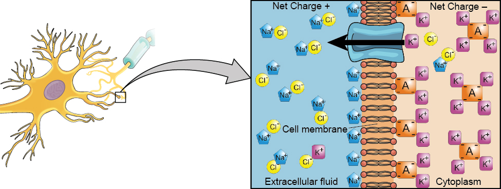
Figure 3.10図 3.10
At resting potential, Na+ (blue pentagons) is more highly concentrated outside the cell in the extracellular fluid (shown in blue), whereas K+ (purple squares) is more highly concentrated near the membrane in the cytoplasm or intracellular fluid. Other molecules, such as chloride ions (yellow circles) and negatively charged proteins (brown squares), help contribute to a positive net charge in the extracellular fluid and a negative net charge in the intracellular fluid.
静止電位では、Na+(青い五角形)は細胞の外側の細胞外液(青で示す)により高い濃度で存在し、K+(紫の四角形)は、細胞質すなわち細胞内液の中の、膜の近くにより高い濃度で存在する。塩化物イオン(黄色の円)や負に帯電したタンパク質(茶色の四角形)など、ほかの分子も、細胞外液が正味で正の電荷を、細胞内液が正味で負の電荷を帯びることに寄与している。
From this resting potential state, the neuron receives a signal and its state changes abruptly (Figure 3.11). When a neuron receives signals at the dendrites—due to neurotransmitters from an adjacent neuron binding to its receptors—small pores, or gates, open on the neuronal membrane, allowing Na+ ions, propelled by both charge and concentration differences, to move into the cell. With this influx of positive ions, the internal charge of the cell becomes more positive. If that charge reaches a certain level, called the threshold of excitation, the neuron becomes active and the action potential begins.
英語のヒント
この静止電位の状態から、ニューロンは信号を受け取り、その状態は急激に変化する(図3.11)。隣接するニューロンからの神経伝達物質が受容体に結合することによって、ニューロンが樹状突起で信号を受け取ると、ニューロンの膜にある小さな孔、つまりゲートが開く。これにより、電荷の差と濃度の差の両方に押されたNa+イオンが、細胞の中へ移動できるようになる。こうして正のイオンが流入すると、細胞の内部の電荷はより正になる。その電荷が、興奮の閾値と呼ばれる一定の水準に達すると、ニューロンは活動状態になり、活動電位が始まる。
Many additional pores open, causing a massive influx of Na+ ions and a huge positive spike in the membrane potential, the peak action potential. At the peak of the spike, the sodium gates close and the potassium gates open. As positively charged potassium ions leave, the cell quickly begins repolarization. At first, it hyperpolarizes, becoming slightly more negative than the resting potential, and then it levels off, returning to the resting potential.
英語のヒント
さらに多くの孔が開き、Na+イオンが大量に流入して、膜電位に大きな正のスパイク、すなわち活動電位のピークが生じる。スパイクの頂点で、ナトリウムのゲートは閉じ、カリウムのゲートが開く。正の電荷を帯びたカリウムイオンが出ていくにつれて、細胞はすばやく再分極を始める。最初は過分極して静止電位よりわずかに負になり、その後は落ち着いて静止電位に戻る。
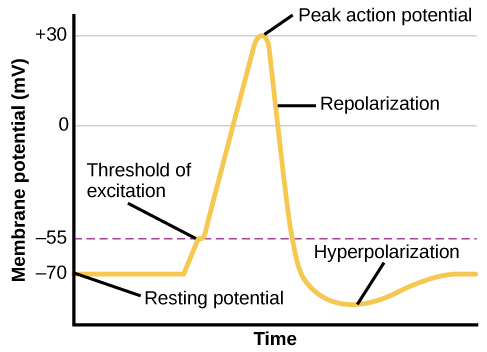
Figure 3.11図 3.11
During the action potential, the electrical charge across the membrane changes dramatically.
活動電位のあいだ、膜をはさんだ電荷は劇的に変化する。
This positive spike constitutes the action potential: the electrical signal that typically moves from the cell body down the axon to the axon terminals. The electrical signal moves down the axon with the impulses jumping in a leapfrog fashion between the Nodes of Ranvier. The Nodes of Ranvier are natural gaps in the myelin sheath. At each point, some of the sodium ions that enter the cell diffuse to the next section of the axon, raising the charge past the threshold of excitation and triggering a new influx of sodium ions. The action potential moves all the way down the axon in this fashion until reaching the terminal buttons.
英語のヒント
この正のスパイクが活動電位にあたる。活動電位とは、ふつう細胞体から軸索を下って軸索終末へと伝わる電気信号である。電気信号は、インパルスがランヴィエ絞輪のあいだを馬跳びのように飛び移りながら、軸索を下っていく。ランヴィエ絞輪は、髄鞘にもともとある隙間である。それぞれの地点で、細胞内に入ったナトリウムイオンの一部が軸索の次の区間へ拡散し、電荷を興奮の閾値より上に押し上げて、ナトリウムイオンの新たな流入を引き起こす。活動電位はこのようにして、終末ボタンに達するまで軸索をずっと下っていく。
The action potential is an all-or-none phenomenon. In simple terms, this means that an incoming signal from another neuron is either sufficient or insufficient to reach the threshold of excitation. There is no in-between, and there is no turning off an action potential once it starts. Think of it like sending an email or a text message. You can think about sending it all you want, but the message is not sent until you hit the send button. Furthermore, once you send the message, there is no stopping it.
英語のヒント
活動電位は全か無かの現象である。簡単に言えば、これは、ほかのニューロンから入ってくる信号が、興奮の閾値に達するのに十分であるか不十分であるかのどちらかだ、ということである。その中間はなく、いったん始まった活動電位を止めることもできない。メールやテキストメッセージを送ることにたとえて考えてみよう。送ろうかと好きなだけ考えていてもよいが、送信ボタンを押すまでメッセージは送られない。さらに、いったんメッセージを送ってしまえば、それを止めることはできない。
Because it is all or none, the action potential is recreated, or propagated, at its full strength at every point along the axon. Much like the lit fuse of a firecracker, it does not fade away as it travels down the axon. It is this all-or-none property that explains the fact that your brain perceives an injury to a distant body part like your toe as equally painful as one to your nose.
英語のヒント
全か無かであるため、活動電位は軸索に沿ったあらゆる地点で、最大の強さのまま再生される、つまり伝播される。火のついた爆竹の導火線とよく似て、活動電位は軸索を下っていくあいだに弱まって消えてしまうことはない。脳が、つま先のような遠く離れた体の部位のけがを、鼻のけがと同じくらい痛いと知覚するという事実を説明するのは、まさにこの全か無かの性質である。
As noted earlier, when the action potential arrives at the terminal button, the synaptic vesicles release their neurotransmitters into the synaptic cleft. The neurotransmitters travel across the synapse and bind to receptors on the dendrites of the adjacent neuron, and the process repeats itself in the new neuron (assuming the signal is sufficiently strong to trigger an action potential). Once the signal is delivered, excess neurotransmitters in the synaptic cleft drift away, are broken down into inactive fragments, or are reabsorbed in a process known as reuptake. Reuptake involves the neurotransmitter being pumped back into the neuron that released it, in order to clear the synapse (Figure 3.12). Clearing the synapse serves both to provide a clear “on” and “off” state between signals and to regulate the production of neurotransmitter (full synaptic vesicles provide signals that no additional neurotransmitters need to be produced).
英語のヒント
前に述べたように、活動電位が終末ボタンに到達すると、シナプス小胞は神経伝達物質をシナプス間隙に放出する。神経伝達物質はシナプスを渡り、隣接するニューロンの樹状突起にある受容体に結合する。そして、新しいニューロンで同じ過程が繰り返される(ただし、信号が活動電位を引き起こすのに十分な強さであればの話である)。信号が伝えられると、シナプス間隙に残った余分な神経伝達物質は、漂って離れていくか、不活性な断片に分解されるか、再取り込みと呼ばれる過程で再吸収される。再取り込みでは、シナプスを空にするために、神経伝達物質が、それを放出したニューロンへとポンプで送り戻される(図3.12)。シナプスを空にすることには、信号と信号のあいだに明確な「オン」と「オフ」の状態をつくる働きと、神経伝達物質の生産を調節する働きの両方がある(シナプス小胞が満杯であれば、それ以上神経伝達物質をつくる必要はないという信号になる)。
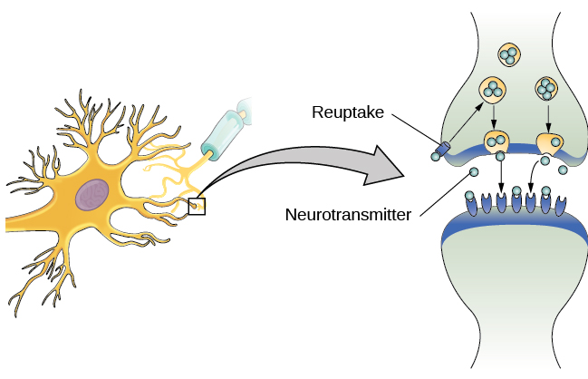


Figure 3.12図 3.12
Reuptake involves moving a neurotransmitter from the synapse back into the axon terminal from which it was released.
再取り込みでは、神経伝達物質が、シナプスから、それを放出した軸索終末へと戻される。
Neuronal communication is often referred to as an electrochemical event. The movement of the action potential down the length of the axon is an electrical event, and movement of the neurotransmitter across the synaptic space represents the chemical portion of the process. However, there are some specialized connections between neurons that are entirely electrical. In such cases, the neurons are said to communicate via an electrical synapse. In these cases, two neurons physically connect to one another via gap junctions, which allows the current from one cell to pass into the next. There are far fewer electrical synapses in the brain, but those that do exist are much faster than the chemical synapses that have been described above (Connors & Long, 2004).
英語のヒント
ニューロンの情報伝達は、しばしば電気化学的な事象と呼ばれる。活動電位が軸索の長さに沿って下っていく動きは電気的な事象であり、神経伝達物質がシナプスの空間を渡っていく動きは、この過程の化学的な部分にあたる。しかし、ニューロンのあいだには、完全に電気的な特殊なつながりもある。そのような場合、ニューロンは電気シナプスを介して情報をやりとりするといわれる。この場合、二つのニューロンはギャップ結合によって物理的に互いにつながっており、これによって一方の細胞の電流が次の細胞へ流れ込むことができる。脳の中の電気シナプスの数ははるかに少ないが、実際に存在するものは、上で述べてきた化学シナプスよりもずっと速い(Connors & Long, 2004)。